Laura Gutiérrez-Miranda
@lgutimiranda.bsky.social
510 followers 680 following 21 posts
Tiny scientist glued to a microscope 🔬. Postdoc at @lymphaticslab.bsky.social Uppsala. Former @weizmann @dkfz.bsky.social alumna #zebrafish #vascularbiology #lymphatics #marrow #microscopy 🧬✂️
Posts Media Videos Starter Packs
Reposted by Laura Gutiérrez-Miranda
janegoodallcan.bsky.social
Dr. Jane made an indelible mark on our understanding of chimpanzees and other species, and also of humankind and the environments we all share.

She inspired curiosity, hope, and compassion in countless people, and paved the way for many others.

#ThankYouJane #RememberingJane

Photo: Marko Zlousic
Reposted by Laura Gutiérrez-Miranda
jem.org
Bassi et al. @karinayaniv.bsky.social @weizmanninstitute.bsky.social use #zebrafish to help identify new treatments for a severe lymphatic malformation. rupress.org/jem/article/...

#CardiovascularBiology #HumanDiseaseGenetics
Reposted by Laura Gutiérrez-Miranda
kevinthiessen.bsky.social
My Bioinformatics Bootcamp turned blog! Enjoy this collection of free #zebrafish resources, now posted on @the-node.bsky.social. While not comprehensive, it gives an assortment of cools tools available to researchers. Feedback would be appreciated! thenode.biologists.com/bioinformati...
lgutimiranda.bsky.social
And just like that we get another #zebrafish surprise !
@pina85.bsky.social work introduces the Inferior Vena Cava vascular remodelling in zebrafish 🦓🐟
Congratulations to all the team for this beautiful story 👏🏻🍾
pina85.bsky.social
I'm thrilled to announce that my #phd work is now out in #biorxiv. Check it out here! I will be happy to have your feedback! www.biorxiv.org/content/10.1... Thank you to @karinayaniv.bsky.social, Rudra N. Das for the menthorship and all authors.
In this work we discovered that during metamophosis there is a remodeling of Zebrafish Cardinal Vein followed by a replacement with the Inferior Vena Cava. This process mirrors the Human Inferior Vena Cava development, demostrating a  conserved mechanism  through  the mammals.  In this panel all the steps of Inferior Vena Cava formation (blood vascular marker, kdrl in green).
lgutimiranda.bsky.social
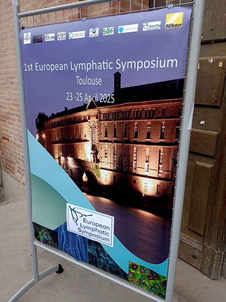
And that is a wrap! Great success of the first European #Lymphatic Symposium
Next one: Budapest 2027
Reposted by Laura Gutiérrez-Miranda
lymphaticslab.bsky.social
🤩 Excited to share our protocol for computationally averaging confocal images of zebrafish lymphatic vessels to identify phenotypes! 🐟🔬 Check it out in Star Protocols 👉 star-protocols.cell.com/protocols/4030

#Zebrafish #Microscopy #StarProtocols
lgutimiranda.bsky.social
Not sure exactly what is going on but I have also received this message today at times where traffic should be rather low 😅
So just to be on the safe side here...Does anybody know if there is a mirror page or alternatives in case one of these days NCBI page is gone? 💨
kaplan.bio
Matias @kaplan.bio · Feb 6
How long does PubMed have? How long will BLAST be up? Genome browser? We have the funding to keep these tools up, we have the expertise, we have the people.

Loss of these tools would cost billions to replace by the private sector and academia. Inefficient destruction with no upside
PubMed site with “Some NLM-NCBI services and products are experiencing heavy traffic, which may affect performance and availability. We apologize for the inconvenience and appreciate your patience. For assistance, please contact our Help Desk at info@ncbi.nlm.nih.gov.”
lgutimiranda.bsky.social
Same here! We can only wait and cross fingers for Rita Fior's lab and all the team at Champalimaud 🤞🏻
lgutimiranda.bsky.social
The first oncology clinical trial using #zebrafish avatars will start this month in Portugal. It is definitely not an easy task. But if it works, it will be a great predictive tool 💪🏻 Good luck!
www.science.org/content/arti...
Fish implanted with tumor cells could help oncologists quickly personalize cancer treatments
The first clinical trial of zebrafish embryos acting as cancer “avatars” will start soon
www.science.org
Reposted by Laura Gutiérrez-Miranda
jcellsci.bsky.social
Aanandita Kothurkar, @colinchulab.bsky.social, Ryan MacDonald and colleagues @ucleye.bsky.social apply IBEX to simultaneously identify all the major retinal cell types.
Highlight: journals.biologists.com/jcs/article/...
Article: journals.biologists.com/jcs/article/...
Figure showing that IBEX can be used to simultaneously label all major retinal types in zebrafish embryos, including confocal images and schematics indicating the labelled cell types. The confocal images show zebrafish retinas labelled with DAPI and 11 different markers: Ribeye-A (dark blue), carbonic anhydrase (CA1, purple), glutamine synthetase (GS, red), PKC-β (green), Zrf-1 (cyan), HuC/D (orange), GFP transgene (yellow), calbindin (CalB, magenta), peripherin-2 (PRPH2, light blue), GNAT2 (orange) and RLBP1 (pink).
Reposted by Laura Gutiérrez-Miranda
biorxivpreprint.bsky.social
Lineage labeling with zebrafish hand2 Cre and CreERT2 recombinase CRISPR knock-ins https://www.biorxiv.org/content/10.1101/2024.12.04.626907v1
Reposted by Laura Gutiérrez-Miranda
zebrafishrock.bsky.social
Help support ZFIN and other MODs by signing this open letter 🙏 🧪
alliancegenome.bsky.social
The Global Biodata Coalition has an open letter campaign to show support for sustainable funding for biodata resources. Please take a minute to read and decide if you want to sign globalbiodata.org/open-letter-...
#biocuration #bioinformatics
OPEN LETTER CAMPAIGN - Global Biodata Coalition
Careers at the Global Biodata Coalition.
globalbiodata.org
Reposted by Laura Gutiérrez-Miranda
dev-journal.bsky.social
Differential vegfc expression dictates lymphatic response during zebrafish heart development and regeneration

Read this Research Article by Sierra Duca, Yu Xia, Michael R. M. Harrison, Jingli Cao and colleagues:
https://buff.ly/49hhwIf
Figure 1 (A) Larval expression of vegfc:EGFP (green) and kdrl:DsRed (magenta) lines in a 2 dpf live fish. Scale bar: 200 µm. (B) Confocal images showing vegfc:EGFP (green) and kdrl:DsRed (magenta) expression in the trunk at 2 dpf. A single-channel image of vegfc:EGFP expression is shown in grayscale on the right. Scale bar: 100 µm. (C,D) Whole-mount images (maximum projections of z-stacks) of the ventricular surface showing expression of vegfc:EGFP (green) and flt1:tdTomato (magenta) at 5 wpf (C) or 8-9 wpf (D). Single-channel images of vegfc:EGFP expression are shown in grayscale on the right. The framed regions are enlarged beneath. The arrows indicate EGFP+tdTpmato+ arteries. Scale bars: 100 µm.
lgutimiranda.bsky.social
Amazing work really, congratulations!!
lgutimiranda.bsky.social
New study on clonal fate during mouse #embryogenesis using in vivo lineage tracing+ deep learning 🤯👏🏻👏🏻
adameykolab.bsky.social
(1) How can we build complex structures from a limited set of progenitors and cell types? In our new preprint, we describe patterns of cell fate decisions during early development and (in some cases) identify molecular correlates of different behaviors.

www.biorxiv.org/content/10.1...
Unbiased profiling of multipotency landscapes reveals spatial modulators of clonal fate biases
Embryogenesis is commonly viewed through a tree model of cell differentiation, which does not adequately represent the spatiotemporal modulation of cell multipotency underlying morphogenesis. Here we ...
www.biorxiv.org
Reposted by Laura Gutiérrez-Miranda
vishalmkrishnan.bsky.social
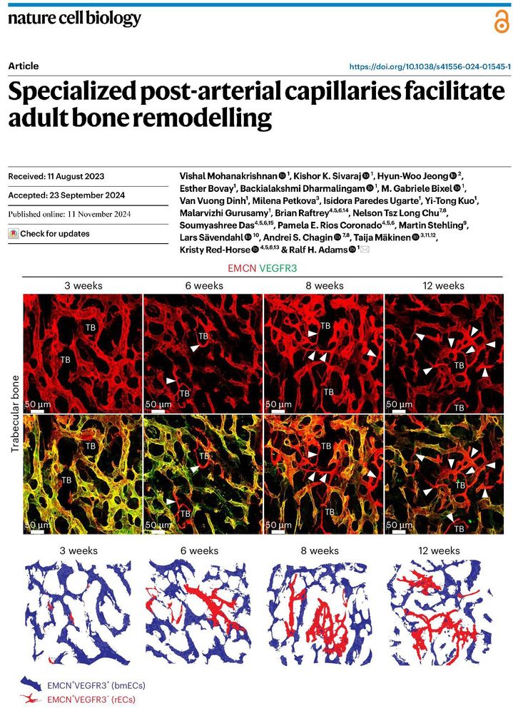
Bone remodeling is traditionally driven by osteoblasts and osteoclasts, but our study reveals a third essential player: "Type R capillaries". 🦴🔬😍

Excited to share my PhD work from Ralf H. Adams lab, now published in @natureportfolio.bsky.social Nature Cell Biology.

www.nature.com/articles/s41...
lgutimiranda.bsky.social
I would like to be added please 🙋🏻‍♀️