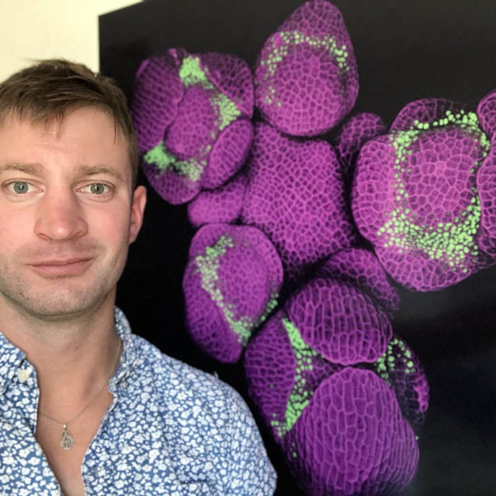
Nat Prunet
@nat-prunet.bsky.social
910 followers 1.2K following 140 posts
Microscopic farmer 🔬🌺 Microscopy core director @UNC Chapel Hill Former plant developmental biologist
Posts Media Videos Starter Packs
nat-prunet.bsky.social
😍
stevengawoski.bsky.social
Humus Sheltered Detritivores 5/2020 22 X 36 inches [55.9 X 91.4 cm] Graphite on Paper #sciart #gawoski #arthropod
Reposted by Nat Prunet
brandy-syglass.bsky.social
👀 #3DThursday: Cross your eyes to see this super res pollen dataset in 3D! Good old fashioned stereo pairs in #syGlass bringing science to life. 🌿✨

🔬: Zeiss LSM 980 with Airyscan 2
🥼: Nat Prunet, @nat-prunet.bsky.social
🏛️: UNC Biological Microscopy Core: tarheels.live/biologymicro...
🎬: syGlass.io
nat-prunet.bsky.social
I played a lot with these guys ha!
Reposted by Nat Prunet
daniellebeckman.bsky.social
For students: how can we quantify neurodegeneration using #microscopy? One way is to use a marker like 🟡 below (SMI32). It identifies a population of pyramidal neurons that is lost early in neurodegenerative diseases like Alzheimer and Parkinson. And after some viral infections 👀👀
#Neuroscience
Pyramidal neurons microscopy image
Reposted by Nat Prunet
the-node.bsky.social
Congratulations to the winners of our image competition with the @mblscience.bsky.social Embryology course. The winner of the Editor’s choice is an image of a anole lizard eye, acquired by Arthur Boutillon @campaslab.bsky.social.
thenode.biologists.com/results-from...
#devbio 🔬🎉
Development cover: Embryonic eye of an anole lizard stained for nuclei (DAPI, blue) and F-actin (Phalloidin, orange), imaged by spinning disc confocal microscopy and processed using ImageJ. The image was acquired at the 2025 MBL Embryology course by Arthur Boutillon and was the Editor's choice from the Node image competition.
Reposted by Nat Prunet
mbfbioscience.bsky.social
🧠🎨 Got brain art?
Submit to our NeuroArt Monthly Image Contest for a chance to be featured + win prizes!

Microscope images, digital renderings, creative interpretations—we want it all.
Let’s celebrate the art in science 🧬✨

🔗 neuroart.com/may-image-co...

#NeuroArt #SciArt #Neuroscience #BrainArt
Reposted by Nat Prunet
nickdesnoyer.bsky.social
Friday Flower 005: Arabidopsis thaliana 🌱✨

A tiny flower, but a giant in research, thale cress enabled our understanding of flower development through the "ABC Model".

With this knowledge, their flowers can be essentially reprogrammed to produce micro-roses 🧬🌹
nat-prunet.bsky.social
shouldn't it be Se Pe Pe Pe when class B genes expand to the center?
Reposted by Nat Prunet
bioimagingna.bsky.social
#HappyFluorescenceFriday!

#microscopycommunity- want to learn open source image analysis or share your knowledge to help others? We’ve got a FREE virtual workshop Nov 17-19! Now accepting workshop session applications!

Learn more & sign up: buff.ly/esGIotD
Reposted by Nat Prunet
minouette.bsky.social
Day 19 #SciArtSeptember 🧪🐡prompt depths: The vampire squid (Vampyroteuthis infernalis), literally the ‘vampire squid from hell’ is a small deep sea cephalopod found throughout temperate and tropical oceans with 2 long retractile filaments, located between the first two pairs of arms on its dorsal 🧵
This is a hand-carved and hand-printed lino block print on delicate ivory Japanese mulberry paper, 9.25” x 12.5” of a vampire squid (Vampyroteuthis infernalis). The ‘vampire squid from hell’ is a small deep sea cephalopod found throughout temperate and tropical oceans with two long retractile filaments, located between the first two pairs of arms on its dorsal side, unlike either octopuses or squids. The squid is upside down with purple head and yellow tent like areas between its legs. The filaments arc around above against the blue background.
nat-prunet.bsky.social
Hey #pollen people! @theobrominated.bsky.social would like to know which species these mixed pollen grains are from, and I admit except for pine and lily, I'm not sure. Would anyone be able to help?

#botany #plantscience #bioimaging #microscopy
nat-prunet.bsky.social
Congrats! Beautiful microscopy too!
Reposted by Nat Prunet
mathpreu.bsky.social
14🧵
Gills offer a powerful model to study :
🩸 Angiogenesis
💨 Blood flow & pressure
🧠 Neural crest stem cells
🌿 Branching morphogenesis — all in vivo.

Our work provides a detailed framework to explore these complex processes.
Reposted by Nat Prunet
bioinformer.bsky.social
This is epic! @officialatcc.bsky.social announced the winners of the 2025 Photo contest! They look amazing! Check the Alt text and follow the link for tons more. www.atcc.org/about-us/upc...

I love this! Please reshare! #microscopy #biology #research #science #cellbiology #microbiology #biotech 🧪🧬
2025 Most Popular Image Award for Microbiology
Bouquet of Trebouxia
Photographer - Sandra Story, NUBAD, LLC
Trebouxia (ATCC 30426) is a free-living green alga that often forms a symbiotic relationship with certain fungi. Trebouxia has a complex life cycle showing many cellular forms including motile. 2025 Most Popular Image Award for Cell Biology
Mitosis in Color: A Super-Resolution View
Photographer - Avishkar Sawant, Vanderbilt University
Super-resolution image of a fixed hTERT-RPE1 cell (ATCC CRL-4000) undergoing mitosis, highlighting the intricate organization of cytoskeletal networks. DNA (cyan), vimentin (depth color-coded), microtubules (red), and actin (magenta) were visualized using NSPARC. The image is presented as a maximum intensity projection of NSPARC stacks. 2025 ATCC Excellence Image Award for Microbiology
Microbial Minimalism
Photographer - Mindy Engevik, Medical University of South Carolina
This is an SEM image of Lactobacillus reuteri (ATCC 6475). 2025 ATCC Excellence Image Award for Microbiology
The Butterfly Effect
Photographer - Sandra Story, NUBAD, LLC
Klebsiella pneumoniae (ATCC 13883) and Bacillus subtilis (ATCC 6501). Fluorescence microscopy composite image of a mixture of bacteria that can be representative of a chronic poly-bacterial infection. Large masses of cells are Klebsiella pneumoniae exposed to a high dose of ertapenem causing the cells to elongate 10-50 times their normal size. Long rigid filaments are Bacillus subtilis common in soil, while Klebsiella pneumoniae are part of the normal microbial flora of the human lower digestive tract. Mixed genera of bacteria can coexist as poly-bacterial biofilms which supports exchange of drug resistance determinants via inter-species and inter-genus horizontal gene transfer. The biofilm-mode of growth supports other synergistic interactions that enhance virulence and promote resistance to antimicrobials resulting in difficult to treat chronic infections. Bacteria were cocultured in nutrient broth exposed to penicillin and incubated at 37 degrees Celsius to allow biofilm development. Samples of the biofilm were stained with three fluorescent dyes FM-64 (red, membrane stain), DAPI (blue, nucleic acid stain for both live and dead cells) and SYTOX Green (green, nucleic acid stain for dead cells) and then spotted onto an agarose nutrient pad sandwiched between a microscope slide and coverslip. The sample was viewed using a Leica DMi8 and illuminated using Texas red, DAPI and GFP filter sets. Images were taken with a K5 microscope camera. Scale bar 20 uM, 1000X magnification.
Reposted by Nat Prunet
france-bioimaging.bsky.social
We are thrilled to announce that our Image Contest is back! 🔬🏆

Submit your best images & try to win your registration fees for one 2026 #microscopy related event.
📆Before November 14th, 2025

Info & entries👉 france-bioimaging.org/fbi-special-...

We are all eager to see your work!
Reposted by Nat Prunet
minouette.bsky.social
I’m making more cyanotypes on handmade paper with a deckle edge:

Fern and wild carrot flower 🐡

minouette.etsy.com/listing/4369...
As described this is a cyanotype on handmade paper with a deckle edge with an image of a fern leaf and a wild carrot flower
Reposted by Nat Prunet
nesmicroscopy.bsky.social
Announcing one of our Keynote Speakers, Elizabeth Engle, for #MicroscopyMonday! Please join us in November at Brandeis!

🔬: 3i Light Sheet
🐭: Dharmendra Puri, Boston Children's Hospital
🏛️: Harvard Medical School's MicRoN facility
🎬: syGlass.io

#NESM #LightSheetMicroscopy #3DImaging #TissueClearing
Reposted by Nat Prunet
jamesdmanton.bsky.social
We present a simple method to easily increase the imageable depth of an expansion microscopy gel on a typical inverted microscope ten-fold, using some carefully placed FEP film and a water dipping objective lens:
A comparison of imaging an expansion microscopy hydrogel with a water dipping objective lens, using either eye gel as an immersion medium or a combination of Immersol W, water and a thin FEP film. In both cases, the sample is supported by a thin FEP film rather than a coverglass.
nat-prunet.bsky.social
😍
liapas.bsky.social
#SciArtSeptember Day 15: spawning • My winter night mitosis sampler (2024) contains two meanings of spawning: it's a human reproductive cell, creating our spawn, and the cytoplasm looks a bit like spawning fish. #SciArt #embroidery
Two reproductive cells dividing embroidered on white linen. The edges of the dividing egg cells and the nucleus membranes are in light grey split stitch. A circle of light purple stem stitch is the cell membrane. The corona radiata is in dark navy zig zag chain stitch. The nucleolus of each cell is in navy bullion stitch. The polar bodies are stitched in burgundy single strand back stitch. The cytoplasm is stitched in variegated blue/purple thread in a mix of seed stitch and French knots. Lia’s initials are stitched in white whipped back stitch.
nat-prunet.bsky.social
This is really cool!
radinbio.bsky.social
Do you know what happens when you touch a carnivorous sundew plant?
If the touch is strong and large enough, a cytosolic calcium wave will spread from the site of touch throughout the whole plant, but if you only touch one tentacle (see post below), the calcium wave will be local and less intense.
Reposted by Nat Prunet
peiferlabunc.bsky.social
If you are interested in the latest advances in Imaging Cell Dynamics, check out the 2026 @jcellsci.bsky.social Meeting. These small meetings are a terrific way to meet key people in the field and get feedback on your own work. Highly recommended!
www.biologists.com/meetings/jcs...
maging Cell Dynamics
11 - 14 May 2026, Montanyà Hotel, Catalonia, Spain

Organisers: Francesca Bottanelli, Guillaume Jacquemet, Michael Way and Giulia Zanetti

Dear colleague,

Following on from our successful meeting in 2023, we are delighted to announce that registration is now open for the second iteration of our Imaging Cell Dynamics meeting, which will be held in May 2026. This meeting will provide a unique opportunity to bring together experts working at the interface between cell biology and imaging, with a focus on researchers who use advanced light and electron microscopy approaches, and novel image analysis tools.

The meeting will feature talks from an outstanding set of invited speakers with opportunities for short talks and poster presentations.

We are delighted to showcase the rapid and exciting progress in this field and to nurture this growing community.

We hope you can join us.

On behalf of the organising committee,
Michael Way
Editor-in-Chief, Journal of Cell Science Organisers and speakers
Francesca Bottanelli Freie Universität Berlin, Germany
Guillaume Jacquemet Åbo Akademi University, Finland
Giulia Zanetti The Francis Crick Institute, UK
Michael Way The Francis Crick Institute, UK

Ori Avinoam Weizmann Institute of Science, Israel
Sarah Cohen UNC-Chapel Hill, USA
Emmanuel Derivery MRC Laboratory of Molecular Biology, UK
Lea Dietrich Max Planck Institute for Brain Research, Germany
Ben Engel University of Basel, Switzerland
Marco Fritzsche University of Oxford, UK
Gerry Hammond University of Pittsburgh, USA
Virginie Hamel University of Geneva, Switzerland
Sven Klumpe Research Institute of Molecular Pathology, Austria
Christophe Leterrier CNRS-Aix Marseille University, France
Dong Li Tsinghua University, China
Taija Mäkinen Uppsala University, Sweden
Jonas Ries Max Perutz Labs Vienna, Austria
Katja Röper MRC Laboratory of Molecular Biology, UK
Loic Royer Chan Zuckerberg Biohub San Francisco, USA
Ilaria Testa KTH Royal Institute of Technology, Sweden
Aubrey Weigel Howard Hughes Medical Institute Janelia Research Campus, USA
nat-prunet.bsky.social
at least this one is not dreaming of eating my persimmons
Reposted by Nat Prunet
healthcare.nikon.com
📄 🌟 Researchers introduce HAK-actin, an actin label compatible with post-labeling in #SuperResolution ultrastructure expansion #Microscopy #ExM. The authors demonstrate its use in cells expanded up to 16-fold (figure).

🔗 View the preprint article: bit.ly/47xC8NJ

#Microscope #Imaging #Fluorescence
Reposted by Nat Prunet
peiferlabunc.bsky.social
OK--fluorescent protein friends. We want an antibody to TagRFP that works in immunofluorescence. Any suggestions? Re-post please
nat-prunet.bsky.social
Beautiful work and gorgeous images!
paveltomancak.bsky.social
We are all super happy and proud to see our work on the function and evolution of the #cephalic #furrow published in @nature.com. Let me say a few things about the background and history of this work on the #Evolution_of_Morphogenesis (1/12)