
Liza Sarde
@sardeliza.bsky.social
47 followers
71 following
5 posts
Ph.D student in @TajbakhshLab.bsky.social @pasteur.fr Stem cell biology & Live Imaging
Posts
Media
Videos
Starter Packs
Pinned

Liza Sarde
@sardeliza.bsky.social
· Mar 14

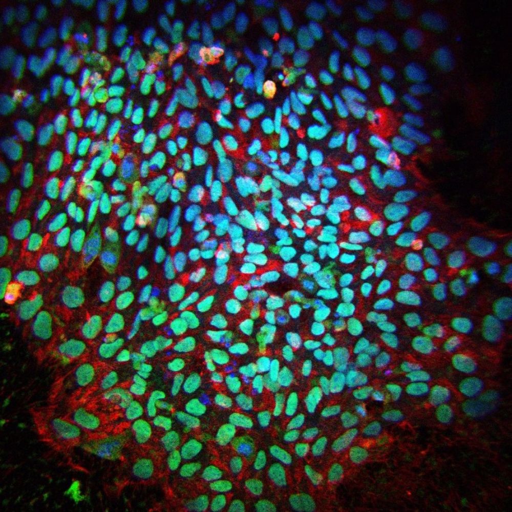
Impaired stem cell migration and divisions in Duchenne Muscular Dystrophy revealed by live imaging
Dysregulation of stem cell properties is a hallmark of many pathologies, but the dynamic behaviour of stem cells in their microenvironment during disease progression remains poorly understood. Using t...
www.biorxiv.org
Liza Sarde
@sardeliza.bsky.social
· Jul 31

Reposted by Liza Sarde

Liza Sarde
@sardeliza.bsky.social
· Mar 15
Reposted by Liza Sarde
Tajbakhsh Lab
@tajbakhshlab.bsky.social
· Mar 14
Liza Sarde
@sardeliza.bsky.social
· Mar 14
Impaired stem cell migration and divisions in Duchenne Muscular Dystrophy revealed by live imaging
Dysregulation of stem cell properties is a hallmark of many pathologies, but the dynamic behaviour of stem cells in their microenvironment during disease progression remains poorly understood. Using t...
www.biorxiv.org
Liza Sarde
@sardeliza.bsky.social
· Mar 14
Impaired stem cell migration and divisions in Duchenne Muscular Dystrophy revealed by live imaging
Dysregulation of stem cell properties is a hallmark of many pathologies, but the dynamic behaviour of stem cells in their microenvironment during disease progression remains poorly understood. Using t...
www.biorxiv.org
Reposted by Liza Sarde

Brendan Evano
@brendan-evano.bsky.social
· Mar 14

Impaired stem cell migration and divisions in Duchenne Muscular Dystrophy revealed by live imaging
Dysregulation of stem cell properties is a hallmark of many pathologies, but the dynamic behaviour of stem cells in their microenvironment during disease progression remains poorly understood. Using t...
www.biorxiv.org
Reposted by Liza Sarde
Brendan Evano
@brendan-evano.bsky.social
· Mar 14
Reposted by Liza Sarde
Brendan Evano
@brendan-evano.bsky.social
· Mar 14
Reposted by Liza Sarde
Reposted by Liza Sarde
Reposted by Liza Sarde

Reposted by Liza Sarde
Reposted by Liza Sarde

Reposted by Liza Sarde
Reposted by Liza Sarde

Reposted by Liza Sarde
Brendan Evano
@brendan-evano.bsky.social
· Mar 14
Liza Sarde
@sardeliza.bsky.social
· Mar 14