Rachel Lee, PhD
@scientistrachel.bsky.social
170 followers 140 following 7 posts
BioImage Data Analyst at AIC Janelia
Posts Media Videos Starter Packs
Reposted by Rachel Lee, PhD
tom-bssnnt.bsky.social
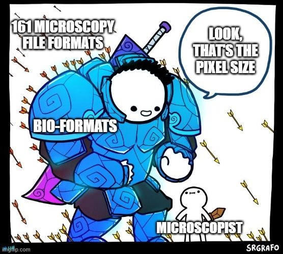
Who does microscopy and has never heard of Bio-Formats? You probably used it anyway
Reposted by Rachel Lee, PhD
aicjanelia.bsky.social
We've enjoyed having Marise and Samara from the lab of @melaniedwhite.bsky.social here to image quail embryos on our MOSAIC system. We wish them safe travels home and happy data analyzing!
A photo of a group of five people standing in a room filled with scientific equipment.
Reposted by Rachel Lee, PhD
joshgondelman.bsky.social
Google search's AI summary is like the loud confident idiot in the room who doesn't always know the answer to your question but HAS TO be the first one to reply to it.
Reposted by Rachel Lee, PhD
melaniedwhite.bsky.social
Today was a super fun first day of imaging @aicjanelia.bsky.social with @vanderspuy.bsky.social and @samarar.bsky.social.
All hail the quail!🧪🔬#DevBio
Reposted by Rachel Lee, PhD
aicjanelia.bsky.social
We are wrapping up a project imaging cancer cell spheroids on our SiMView system today. We wish Elizabeta Belova of @temple.edu happy data analyzing and safe travels home!
A photo of a group of people. Two people are sitting in chairs with a small table between them, while the remaining four people stand behind the chairs. The white wall behind the group is decorated with microscopy images.
Reposted by Rachel Lee, PhD

aicjanelia.bsky.social
Today we're wrapping up a dicty project on our MOSAIC system. We've enjoyed having Yiyan and Saki of the Devreotes lab here and wish them happy data analyzing!
A group of four people stand in front of a microscope system. They are surrounded by scientific equipment, including two large computer monitors.
Reposted by Rachel Lee, PhD
aicjanelia.bsky.social
We're wrapping up a fun three weeks of drosophila imaging on our SiMView microscope with the Mavrakis lab from @institutfresnel.bsky.social Wishing their team safe travels home and happy data analyzing!
A photo of a group of people standing or sitting in front of a white wall.
Reposted by Rachel Lee, PhD
aicjanelia.bsky.social
Just over a week left to apply for this round of the AIC Call for Proposals! Submit at www.aicjanelia.org/apply by Monday, June 16 (5 pm US ET).
aicjanelia.bsky.social
The latest AIC Call for Proposals is now open! Deadline is June 16, 2025. Come use @hhmijanelia.bsky.social imaging technology and expertise for FREE. Visit www.aicjanelia.org/apply for details. Before applying, contact us to schedule a technical consultation!
Apply | aicjanelia
Apply to use the advanced microscopes housed at the AIC.
www.aicjanelia.org
Reposted by Rachel Lee, PhD
blankenshiplab.bsky.social
Back MOSAICing @hhmijanelia.bsky.social... as always a great imaging experience, and they have an open call for apps: www.aicjanelia.org/apply @aicjanelia.bsky.social
Reposted by Rachel Lee, PhD
aicjanelia.bsky.social
Last week we wrapped up an iPALM project with Shannon from the @mrobichaux.bsky.social lab at #WVU. Wishing her luck with her data analysis!
A photo of two women sitting in blue chairs with a small table between them. Behind them on the wall are framed microscopy images.
scientistrachel.bsky.social
April is #CitizenScienceMonth! There is still time to join @scistarter.bsky.social and be part of #OneMillionActsofScience 2025. I had fun observing some trees for the Globe Observer program 🌳
Reposted by Rachel Lee, PhD
cellsandculture.bsky.social
We’ve had an incredible experience being hosted at the AIC at Janelia for iPALM imaging. Tremendous support and expertise.
If your project could benefit from advanced imaging, check out their instruments (FIB-SEM, iPALM, LLSM and others) and get in touch.👇
aicjanelia.bsky.social
The latest AIC Call for Proposals is now open! Deadline is June 16, 2025. Come use @hhmijanelia.bsky.social imaging technology and expertise for FREE. Visit www.aicjanelia.org/apply for details. Before applying, contact us to schedule a technical consultation!
Apply | aicjanelia
Apply to use the advanced microscopes housed at the AIC.
www.aicjanelia.org
scientistrachel.bsky.social
Good advice! 👇
jcellsci.bsky.social
Joanna Pylvänäinen @jwpylvanainen.bsky.social Hanna Grobe and Guillaume Jacquemet @guijacquemet.bsky.social present their practical guidelines for data exploration in quantitative cell biology.
journals.biologists.com/jcs/article/...
#OpenAccess
Here, we show data exploration and visualization for an experiment comparing the number of filopodia in control cells with that in cells treated with siRNA targeting TLNRD1. (A) HUVECs expressing Lifeact–RFP under control or experimental conditions. Cells were fixed, stained with DAPI and imaged using spinning disk confocal microscopy. Representative maximal intensity projections of fields of view are shown. Scale bar: 20 µm. Images were acquired as part of a previously published study (Ball et al., 2024). (B) Schematic of the wide data format, where data relating to the number of filopodia counted in each condition are stored across columns, making data more challenging to manipulate for downstream analyses. (C) Schematic of the tidy data format, where each variable (e.g. condition, filopodia count, repeat) is stored in a single column, and each observation occupies a row. This organization enables flexible data wrangling, statistical analysis and direct input into visualization tools such as box plots and heatmaps. (D) The number of filopodia per GFP-positive cell as shown in A was manually quantified across all conditions (four biological repeats, >60 fields of view per condition). Results are presented as box plots, where horizontal lines mark the median, boxes represent the interquartile range, and vertical whiskers extend to data points within 1.5× the interquartile range above and below the third and first quartiles, respectively. Dot plots showing each biological replicate uniquely color-coded are also presented as part of a SuperPlot (Lord et al., 2020) to visualize variability in the data. Importantly, the tidy data format was required to make this plot. (E) Pairwise condition comparisons using heatmaps. Three mirrored heatmaps, one displaying the effect size (Cohen's d values) and two displaying the statistical significance (P-values from randomization tests with and without Bonferroni correction). The plots in D and E were generated using Plot-Stats.
Reposted by Rachel Lee, PhD
anjanja1024.bsky.social
A new call for proposals is open at our Advanced Imaging Center for imaging on our pre-commercial microscopes. Reach out for a technical consultation and learn how to apply to use the newest technology for free!
aicjanelia.bsky.social
The latest AIC Call for Proposals is now open! Deadline is June 16, 2025. Come use @hhmijanelia.bsky.social imaging technology and expertise for FREE. Visit www.aicjanelia.org/apply for details. Before applying, contact us to schedule a technical consultation!
Apply | aicjanelia
Apply to use the advanced microscopes housed at the AIC.
www.aicjanelia.org
scientistrachel.bsky.social
Applications are open for using @aicjanelia.bsky.social microscopes! Reach out to our team for a free technical consultation to get feedback on your proposal before submission.
aicjanelia.bsky.social
The latest AIC Call for Proposals is now open! Deadline is June 16, 2025. Come use @hhmijanelia.bsky.social imaging technology and expertise for FREE. Visit www.aicjanelia.org/apply for details. Before applying, contact us to schedule a technical consultation!
Apply | aicjanelia
Apply to use the advanced microscopes housed at the AIC.
www.aicjanelia.org
Reposted by Rachel Lee, PhD
aicjanelia.bsky.social
Today we're wrapping up an iPALM project with John Henson (Dickinson College) and Brad Shuster (@nmsu.edu). We've enjoyed having them (and their sea urchins) here at Janelia, and wish them safe travels home!
A photo of a group of people standing together inside a laboratory. There is a tank with sea urchins in the background. A photo of a tank of search urchins.
Reposted by Rachel Lee, PhD
aicjanelia.bsky.social
And the Nejsum Lab (nejsumlab.au.dk) from @au.dk is wrapping imaging our on our MOSAIC system. We wish both groups safe travels home!
A photo of a group of people in front of a display of computer monitors.
Reposted by Rachel Lee, PhD
aicjanelia.bsky.social
This week we're wrapping up two AIC visits at once! @kylejacobs.bsky.social from UCSF was here for a project on our iPALM system.
A photo of a group of people.
Reposted by Rachel Lee, PhD
scopeshifu.bsky.social
🧪Job opening at Janelia @hhmijanelia.bsky.social Integrative Imaging platform for a bioimage analyst. Please repost.
i2janelia.bsky.social
We have an open #opportunity for a BioImage Analyst to join our team! This is an excellent role for a problem solver with an empathetic, collaborative mentality who enjoys partnering with interdisciplinary scientists. Read more and apply here: hhmi.wd1.myworkdayjobs.com/en-US/Extern...
BioImage Analyst
Primary Work Address: 19700 Helix Drive, Ashburn, VA, 20147 Current HHMI Employees, click here to apply via your Workday account. The Howard Hughes Medical Institute’s Janelia Research Campus is a pio...
hhmi.wd1.myworkdayjobs.com
Reposted by Rachel Lee, PhD
aicjanelia.bsky.social
Opportunity to join Integrative Imaging and work with the AIC team on #bioimageanalysis! 👇
i2janelia.bsky.social
We have an open #opportunity for a BioImage Analyst to join our team! This is an excellent role for a problem solver with an empathetic, collaborative mentality who enjoys partnering with interdisciplinary scientists. Read more and apply here: hhmi.wd1.myworkdayjobs.com/en-US/Extern...
BioImage Analyst
Primary Work Address: 19700 Helix Drive, Ashburn, VA, 20147 Current HHMI Employees, click here to apply via your Workday account. The Howard Hughes Medical Institute’s Janelia Research Campus is a pio...
hhmi.wd1.myworkdayjobs.com
scientistrachel.bsky.social
This is an #opportunity to join Integrative Imaging and work with the @aicjanelia.bsky.social team! I enjoy getting to help researchers design and implement #bioimageanalysis approaches for a wide variety of #microscopy projects in this role 🤓
i2janelia.bsky.social
We have an open #opportunity for a BioImage Analyst to join our team! This is an excellent role for a problem solver with an empathetic, collaborative mentality who enjoys partnering with interdisciplinary scientists. Read more and apply here: hhmi.wd1.myworkdayjobs.com/en-US/Extern...
BioImage Analyst
Primary Work Address: 19700 Helix Drive, Ashburn, VA, 20147 Current HHMI Employees, click here to apply via your Workday account. The Howard Hughes Medical Institute’s Janelia Research Campus is a pio...
hhmi.wd1.myworkdayjobs.com
Reposted by Rachel Lee, PhD
aicjanelia.bsky.social
Wishing Alex George and team happy data analyzing now that they've wrapped up their imaging on our MOSAIC system!
A photo of a group of people. One row of people sits on a bench while another row stands behind. Behind the people are large windows revealing a sunny outdoor scene.
Reposted by Rachel Lee, PhD
jytinevez.bsky.social
10 - Featured extension: Mastodon-Blender

Offers a bridge from Mastodon to Blender to create high quality 3D rendering of lineages and cell tracks over time:

(Tracking data of phallusia mammillata embryogenesis by Guignard et a. (2020). doi.org/10.1126/scie...)
Reposted by Rachel Lee, PhD
Thank you for hosting our team and making this project a success! Here is a macrophage playing beach volleyball with IgG-coated beads, courtesy of Tyler Paul, Yoseph Loyd, and @scientistrachel.bsky.social !