


Aurélien Villedieu
@aurelienvilledieu.bsky.social
Postdoc in Jérôme Gros's team | Former PhD student in Yohanns Bellaïche's team | Multi-scale study of tissue morphogenesis | Live-imaging and quantitative biology
Reposted by Aurélien Villedieu

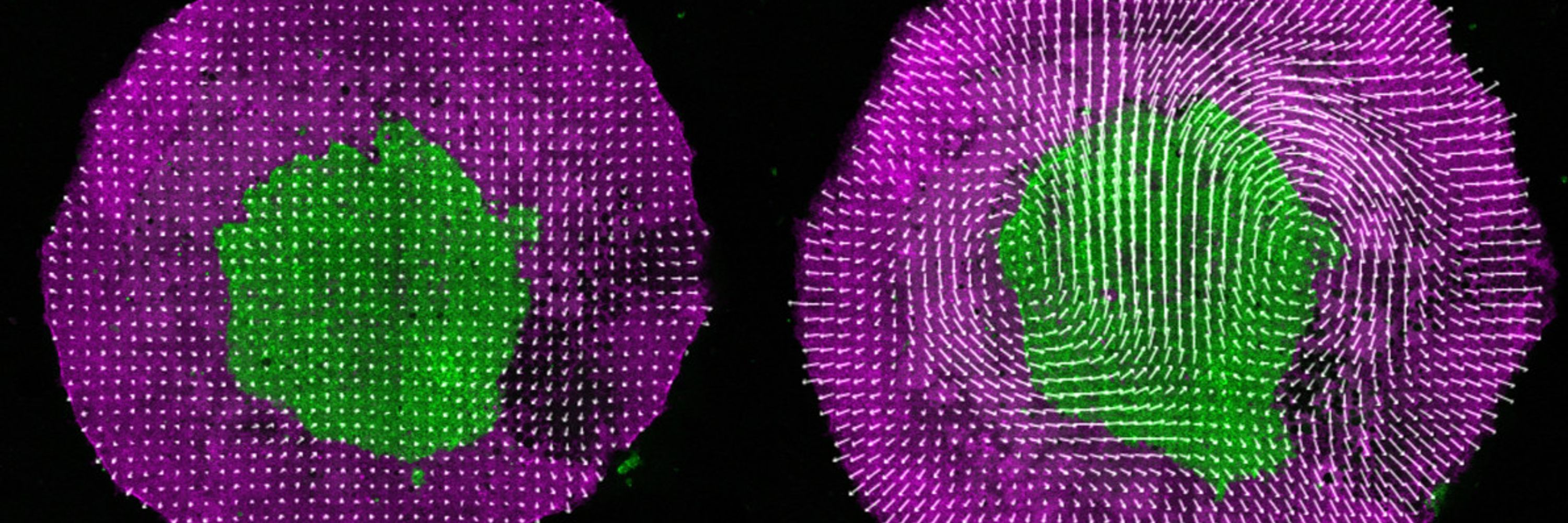
#RésultatScientifique 🔎 | Comment les filaments d'actine s'organisent à l'intérieur des cellules et des tissus vivants
🔬 @institutfresnel.bsky.social 🤝 @univ-amu.fr @centralemed.bsky.social @cnrs.fr
📍 @cnrs-dr12.bsky.social
🔬 @institutfresnel.bsky.social 🤝 @univ-amu.fr @centralemed.bsky.social @cnrs.fr
📍 @cnrs-dr12.bsky.social

Comment les filaments d'actine s'organisent à l'intérieur des cellules et des tissus vivants
En utilisant la microscopie de fluorescence résolue en polarisation, les biologistes et physiciens de l'Institut Fresnel, associés à une équipe internat
www.insis.cnrs.fr
November 26, 2025 at 9:58 AM
#RésultatScientifique 🔎 | Comment les filaments d'actine s'organisent à l'intérieur des cellules et des tissus vivants
🔬 @institutfresnel.bsky.social 🤝 @univ-amu.fr @centralemed.bsky.social @cnrs.fr
📍 @cnrs-dr12.bsky.social
🔬 @institutfresnel.bsky.social 🤝 @univ-amu.fr @centralemed.bsky.social @cnrs.fr
📍 @cnrs-dr12.bsky.social
Reposted by Aurélien Villedieu

Out today. 🙏 again to everyone for this wonderful piece of work, in particular to Aurelie @aurhin.bsky.social Chase @chasebolt.bsky.social and Brent @homeobox.bsky.social. 🙏 also to the Harris lab @fish4walking.bsky.social and @neilshubin.bsky.social @biology-unige.bsky.social @college-de-france.fr

September 17, 2025 at 5:30 PM
Out today. 🙏 again to everyone for this wonderful piece of work, in particular to Aurelie @aurhin.bsky.social Chase @chasebolt.bsky.social and Brent @homeobox.bsky.social. 🙏 also to the Harris lab @fish4walking.bsky.social and @neilshubin.bsky.social @biology-unige.bsky.social @college-de-france.fr
Reposted by Aurélien Villedieu

We are all super happy and proud to see our work on the function and evolution of the #cephalic #furrow published in @nature.com. Let me say a few things about the background and history of this work on the #Evolution_of_Morphogenesis (1/12)

September 4, 2025 at 8:22 AM
We are all super happy and proud to see our work on the function and evolution of the #cephalic #furrow published in @nature.com. Let me say a few things about the background and history of this work on the #Evolution_of_Morphogenesis (1/12)
Reposted by Aurélien Villedieu
Our latest: We developed a chemo-optogenetic system for precise spatiotemporal control of morphogen production. Using dual light + small molecule control of Sonic Hedgehog production, we recapitulated neural tube patterning in vitro & measured spread of Shh
🧵
www.sciencedirect.com/science/arti...
🧵
www.sciencedirect.com/science/arti...

Investigating morphogen and patterning dynamics with optogenetic control of morphogen production
Morphogen gradients provide the patterning cues that instruct cell fate decisions during development. Here, we establish an optogenetic system for the…
www.sciencedirect.com
August 26, 2025 at 8:28 AM
Our latest: We developed a chemo-optogenetic system for precise spatiotemporal control of morphogen production. Using dual light + small molecule control of Sonic Hedgehog production, we recapitulated neural tube patterning in vitro & measured spread of Shh
🧵
www.sciencedirect.com/science/arti...
🧵
www.sciencedirect.com/science/arti...
Reposted by Aurélien Villedieu

Latest paper elifesciences.org/articles/107... closes an important cycle in our efforts to study regeneration: week-long recordings allow us to observe the behaviour of cells during the entire course of regeneration in a crustacean leg – bright objects in movie are fluorescent nuclei of cells. 1/6
August 8, 2025 at 5:39 PM
Latest paper elifesciences.org/articles/107... closes an important cycle in our efforts to study regeneration: week-long recordings allow us to observe the behaviour of cells during the entire course of regeneration in a crustacean leg – bright objects in movie are fluorescent nuclei of cells. 1/6
Reposted by Aurélien Villedieu
Very happy that the first article from my postdoc work in the Tomancak lab is now published @PNAS! www.pnas.org/doi/10.1073/.... We studied the self-organization of actin in aggregates made from Hydra cells. Thread below (1/9)
August 6, 2025 at 1:09 PM
Very happy that the first article from my postdoc work in the Tomancak lab is now published @PNAS! www.pnas.org/doi/10.1073/.... We studied the self-organization of actin in aggregates made from Hydra cells. Thread below (1/9)
Reposted by Aurélien Villedieu

Finally out! 🥳 Our paper showing how a transposable element (TE) insertion can cause developmental phenotypes is now published @natgenet.nature.com 🧬🦠🐁
Below is a brief description of the major findings. Check the full version of the paper for more details: www.nature.com/articles/s41588-025-02248-5
Below is a brief description of the major findings. Check the full version of the paper for more details: www.nature.com/articles/s41588-025-02248-5
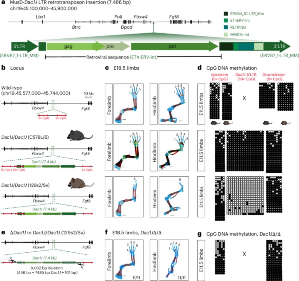
Enhancer adoption by an LTR retrotransposon generates viral-like particles, causing developmental limb phenotypes - Nature Genetics
Activation of an LTR retrotransposon inserted upstream of the Fgf8 gene produces viral-like particles in the mouse developing limb, triggering apoptosis and causing limb malformation. This phenotype c...
www.nature.com
July 9, 2025 at 10:05 AM
Finally out! 🥳 Our paper showing how a transposable element (TE) insertion can cause developmental phenotypes is now published @natgenet.nature.com 🧬🦠🐁
Below is a brief description of the major findings. Check the full version of the paper for more details: www.nature.com/articles/s41588-025-02248-5
Below is a brief description of the major findings. Check the full version of the paper for more details: www.nature.com/articles/s41588-025-02248-5
Reposted by Aurélien Villedieu

If you’ve been following this account closely, you might already know methods to probe mechanics in vitro, but what about in live embryos?
I’m @amichaut.bsky.social, and I’m going to share a few great papers on this aspect of #EpithelialMechanics.
I’m @amichaut.bsky.social, and I’m going to share a few great papers on this aspect of #EpithelialMechanics.
July 20, 2025 at 7:01 AM
If you’ve been following this account closely, you might already know methods to probe mechanics in vitro, but what about in live embryos?
I’m @amichaut.bsky.social, and I’m going to share a few great papers on this aspect of #EpithelialMechanics.
I’m @amichaut.bsky.social, and I’m going to share a few great papers on this aspect of #EpithelialMechanics.
Reposted by Aurélien Villedieu

Here is what I have been up to in the lab of @stephangrill.bsky.social together with @neipel.bsky.social and many others.
Intrigued by avian left right symmetry breaking we found that a tissue-scale active torque drives chiral flow and requires mechanical coupling to the underlying tissue.
Intrigued by avian left right symmetry breaking we found that a tissue-scale active torque drives chiral flow and requires mechanical coupling to the underlying tissue.

(1/10)
I am excited to finally share our work on avian left/right symmetry breaking with you. We reveal a tissue-scale active torque dipole generated at the Hensen’s node, thanks to crazy experiments by Julia @juliapfanzelter.bsky.social and some analysis by me.
doi.org/10.1101/2025...
I am excited to finally share our work on avian left/right symmetry breaking with you. We reveal a tissue-scale active torque dipole generated at the Hensen’s node, thanks to crazy experiments by Julia @juliapfanzelter.bsky.social and some analysis by me.
doi.org/10.1101/2025...

An active torque dipole across tissue layers drives avian left-right symmetry breaking
Unlike in mice, frogs, and fish, left-right (L/R) body axis formation in avian embryos does not arise from the chiral beat of cilia. Instead, a counter-clockwise tissue rotation around Hensen′ node, t...
doi.org
July 20, 2025 at 3:53 PM
Here is what I have been up to in the lab of @stephangrill.bsky.social together with @neipel.bsky.social and many others.
Intrigued by avian left right symmetry breaking we found that a tissue-scale active torque drives chiral flow and requires mechanical coupling to the underlying tissue.
Intrigued by avian left right symmetry breaking we found that a tissue-scale active torque drives chiral flow and requires mechanical coupling to the underlying tissue.
Reposted by Aurélien Villedieu

🏳️🌈Our editor @slefkopoulos.bsky.social discussed with @jzylicz.bsky.social about the importance of celebrating Pride, representation in science, and more! Happy Pride!
Read here: rdcu.be/ewfba
bit.ly/3Uc72TO
Read here: rdcu.be/ewfba
bit.ly/3Uc72TO

Coming out is not a one-time event - Nature Cell Biology
June is the month celebrating Pride in the USA and other countries around the world to honour the Stonewall Uprising of 1969, as well as all progress and current strives claiming equal justice for mem...
bit.ly
July 14, 2025 at 11:36 PM
🏳️🌈Our editor @slefkopoulos.bsky.social discussed with @jzylicz.bsky.social about the importance of celebrating Pride, representation in science, and more! Happy Pride!
Read here: rdcu.be/ewfba
bit.ly/3Uc72TO
Read here: rdcu.be/ewfba
bit.ly/3Uc72TO
Many thanks to Ruoheng Li and @prelights.bsky.social for featuring our preprint!

Seeing and (not) believing! On the role of hypoblast in primitive streak induction.
In her second #preLight, Ruoheng Li covers the #preprint by
@aurelienvilledieu.bsky.social and the @jeromegros.bsky.social's research team.
In her second #preLight, Ruoheng Li covers the #preprint by
@aurelienvilledieu.bsky.social and the @jeromegros.bsky.social's research team.
Live imaging and functional characterization of the avian hypoblast redefine the mechanisms of primitive streak induction - preLights
Seeing and (not) believing the role of hypoblast in primitive streak induction
prelights.biologists.com
July 10, 2025 at 5:08 PM
Many thanks to Ruoheng Li and @prelights.bsky.social for featuring our preprint!
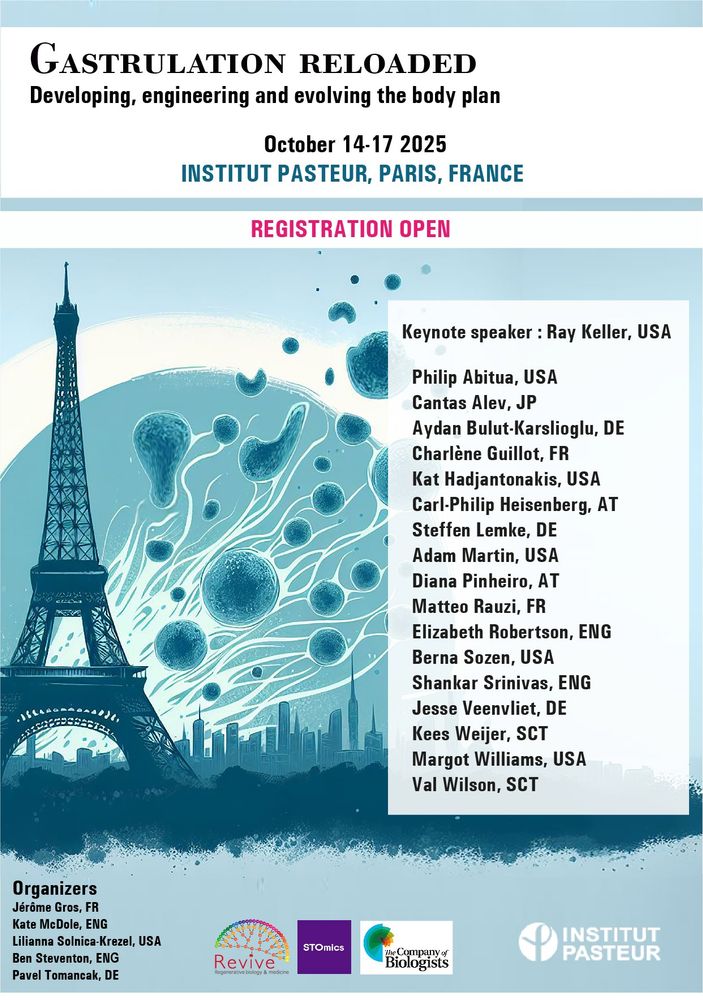
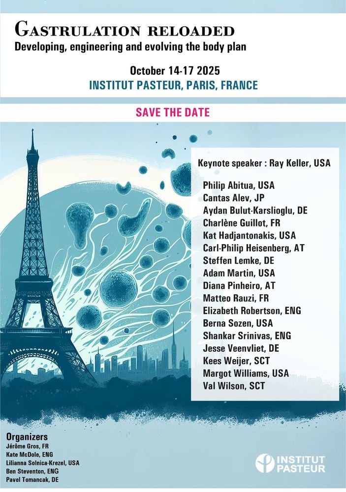
🚨 A very interesting conference on gastrulation coming up soon 🚨 Don't miss the chance to interact with these renowned speakers! Early bird deadline: July 15th
Registration is now open!
Gastrulation Reloaded: Developing, engineering & evolving the body plan
📅 October 14–17, 2025
📍 Paris, France
🗓️ Abstract & early bird deadline: July 15, 2025
www.gastrulation-reloaded.conferences-pasteur.org/home
Exceptional lineup of speakers & many selected talks
Gastrulation Reloaded: Developing, engineering & evolving the body plan
📅 October 14–17, 2025
📍 Paris, France
🗓️ Abstract & early bird deadline: July 15, 2025
www.gastrulation-reloaded.conferences-pasteur.org/home
Exceptional lineup of speakers & many selected talks
July 3, 2025 at 1:25 PM
🚨 A very interesting conference on gastrulation coming up soon 🚨 Don't miss the chance to interact with these renowned speakers! Early bird deadline: July 15th
Reposted by Aurélien Villedieu

🧵Just out !
We reveal how 4 branched epithelia—mammary, lacrimal, salivary & prostate—use a conserved YAP–Notch–p63 circuit to self-organize during development & regeneration.
Here’s the story👇
authors.elsevier.com/c/1lM285Sx5g...
Work done in @frelab.bsky.social at
@institutcurie.bsky.social
We reveal how 4 branched epithelia—mammary, lacrimal, salivary & prostate—use a conserved YAP–Notch–p63 circuit to self-organize during development & regeneration.
Here’s the story👇
authors.elsevier.com/c/1lM285Sx5g...
Work done in @frelab.bsky.social at
@institutcurie.bsky.social
July 1, 2025 at 6:39 AM
🧵Just out !
We reveal how 4 branched epithelia—mammary, lacrimal, salivary & prostate—use a conserved YAP–Notch–p63 circuit to self-organize during development & regeneration.
Here’s the story👇
authors.elsevier.com/c/1lM285Sx5g...
Work done in @frelab.bsky.social at
@institutcurie.bsky.social
We reveal how 4 branched epithelia—mammary, lacrimal, salivary & prostate—use a conserved YAP–Notch–p63 circuit to self-organize during development & regeneration.
Here’s the story👇
authors.elsevier.com/c/1lM285Sx5g...
Work done in @frelab.bsky.social at
@institutcurie.bsky.social
Reposted by Aurélien Villedieu

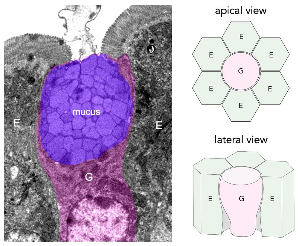
🥳 Thrilled to share our lab's first preprint, led by our talented postdoc Justine Creff! 👩🏻🔬 We tackled a fundamental question: how the epithelium withstands mechanical stress at the interface of cells with distinct geometries and mechanics, such as enterocytes (E) and goblet cells (G) (1/9)
April 6, 2025 at 10:24 PM
🥳 Thrilled to share our lab's first preprint, led by our talented postdoc Justine Creff! 👩🏻🔬 We tackled a fundamental question: how the epithelium withstands mechanical stress at the interface of cells with distinct geometries and mechanics, such as enterocytes (E) and goblet cells (G) (1/9)
Reposted by Aurélien Villedieu

Very happy to share our latest lab publication!
The hard work of @bboumard.bsky.social, Gwenn Le Meur and collaborators!
Cell-type-specific nucleotide sharing through gap junctions impacts sensitivity to replication stress in Drosophila: Developmental Cell
authors.elsevier.com/a/1lDFi_Yv6z...
The hard work of @bboumard.bsky.social, Gwenn Le Meur and collaborators!
Cell-type-specific nucleotide sharing through gap junctions impacts sensitivity to replication stress in Drosophila: Developmental Cell
authors.elsevier.com/a/1lDFi_Yv6z...
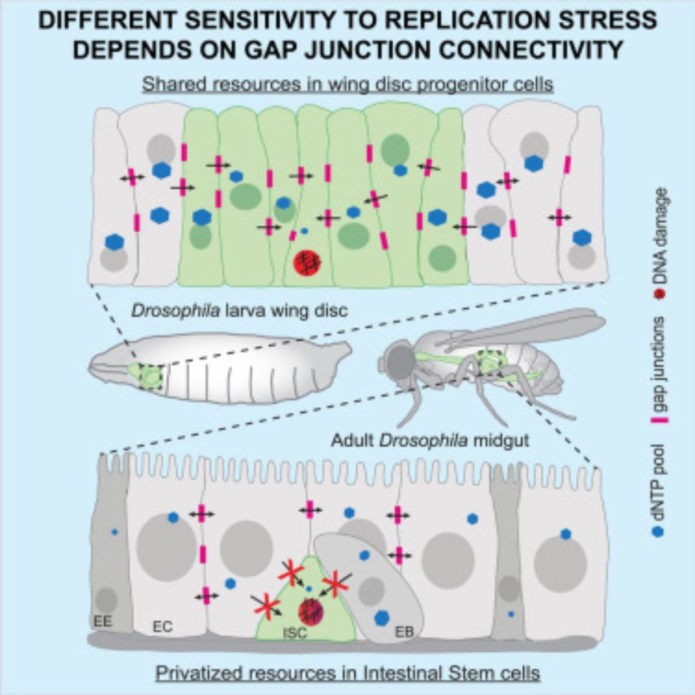
Cell-type-specific nucleotide sharing through gap junctions impacts sensitivity to replication stress in Drosophila
Boumard et al. demonstrate gap-junction-dependent tissue-scale nucleotide sharing,
which impacts cellular sensitivity to perturbation of nucleotide homeostasis and replication
stress. Drosophila wing ...
www.cell.com
June 5, 2025 at 4:47 PM
Very happy to share our latest lab publication!
The hard work of @bboumard.bsky.social, Gwenn Le Meur and collaborators!
Cell-type-specific nucleotide sharing through gap junctions impacts sensitivity to replication stress in Drosophila: Developmental Cell
authors.elsevier.com/a/1lDFi_Yv6z...
The hard work of @bboumard.bsky.social, Gwenn Le Meur and collaborators!
Cell-type-specific nucleotide sharing through gap junctions impacts sensitivity to replication stress in Drosophila: Developmental Cell
authors.elsevier.com/a/1lDFi_Yv6z...
Reposted by Aurélien Villedieu

The effector caspases drive cell death, but their activation can often be survived, so how do cells make this decision? Our new preprint from @levayerr.bsky.social shows that instantaneous caspase activity is important, but past activation has a key role to play!
www.biorxiv.org/content/10.1...
www.biorxiv.org/content/10.1...
May 21, 2025 at 8:52 AM
The effector caspases drive cell death, but their activation can often be survived, so how do cells make this decision? Our new preprint from @levayerr.bsky.social shows that instantaneous caspase activity is important, but past activation has a key role to play!
www.biorxiv.org/content/10.1...
www.biorxiv.org/content/10.1...
🚨New preprint
Glad to share my postdoc work on the role of the hypoblast in primitive streak induction! Discover how live imaging observations led us to revisit the molecular mechanisms governing embryonic axis induction
Thanks to @jeromegros.bsky.social and all co-authors!
doi.org/10.1101/2025...
Glad to share my postdoc work on the role of the hypoblast in primitive streak induction! Discover how live imaging observations led us to revisit the molecular mechanisms governing embryonic axis induction
Thanks to @jeromegros.bsky.social and all co-authors!
doi.org/10.1101/2025...

Live imaging and functional characterization of the avian hypoblast redefine the mechanisms of primitive streak induction
In birds and mammals, the formation of the primitive streak, the hallmark of the primary axis and site of gastrulation, is thought to occur when an anterior displacement of the hypoblast (visceral end...
doi.org
May 19, 2025 at 8:59 AM
🚨New preprint
Glad to share my postdoc work on the role of the hypoblast in primitive streak induction! Discover how live imaging observations led us to revisit the molecular mechanisms governing embryonic axis induction
Thanks to @jeromegros.bsky.social and all co-authors!
doi.org/10.1101/2025...
Glad to share my postdoc work on the role of the hypoblast in primitive streak induction! Discover how live imaging observations led us to revisit the molecular mechanisms governing embryonic axis induction
Thanks to @jeromegros.bsky.social and all co-authors!
doi.org/10.1101/2025...
Reposted by Aurélien Villedieu
🚨📣 New preprint alert!
Excited to share my new postdoc work on direct force and mechanical properties measurements in live embryos!
A great team effort with A. Chamolly (theory), under the supervision of F. Corson and @jeromegros.bsky.social
📄 www.biorxiv.org/content/10.1...
#devbio #mechanics
Excited to share my new postdoc work on direct force and mechanical properties measurements in live embryos!
A great team effort with A. Chamolly (theory), under the supervision of F. Corson and @jeromegros.bsky.social
📄 www.biorxiv.org/content/10.1...
#devbio #mechanics

Direct measurements of active forces and material properties unveil the active mechanics of early embryogenesis
Despite progress in probing tissue mechanics, direct long-term measurements in live embryonic epithelia are lacking. This limits our understanding of amniote embryonic morphogenesis, which takes place...
www.biorxiv.org
May 12, 2025 at 2:01 PM
🚨📣 New preprint alert!
Excited to share my new postdoc work on direct force and mechanical properties measurements in live embryos!
A great team effort with A. Chamolly (theory), under the supervision of F. Corson and @jeromegros.bsky.social
📄 www.biorxiv.org/content/10.1...
#devbio #mechanics
Excited to share my new postdoc work on direct force and mechanical properties measurements in live embryos!
A great team effort with A. Chamolly (theory), under the supervision of F. Corson and @jeromegros.bsky.social
📄 www.biorxiv.org/content/10.1...
#devbio #mechanics
Reposted by Aurélien Villedieu
ICYMI: Commentary on "mechanism" in dev bio
Ozpolat et al argue for diverse approaches to mechanism - from molecular to systems level
journals.biologists.com/dev/article/...
Ozpolat et al argue for diverse approaches to mechanism - from molecular to systems level
journals.biologists.com/dev/article/...
A case for broadening our view of mechanism in developmental biology
ABSTRACT. Developmental biologists can perform studies that describe a phenomenon (descriptive work) and/or explain how the phenomenon works (mechanistic work). There is a prevalent perception that mo...
journals.biologists.com
February 5, 2025 at 10:56 AM
ICYMI: Commentary on "mechanism" in dev bio
Ozpolat et al argue for diverse approaches to mechanism - from molecular to systems level
journals.biologists.com/dev/article/...
Ozpolat et al argue for diverse approaches to mechanism - from molecular to systems level
journals.biologists.com/dev/article/...
Reposted by Aurélien Villedieu
Announcing this upcoming conference:
GASTRULATION RELOADED:
Developing, engineering, and evolving the body plan
Join us as we explore the latest research on gastrulation and axis formation/elongation, featuring common, exotic, and synthetic embryos!
📅 14–17 October 2025
📍 Paris, France
GASTRULATION RELOADED:
Developing, engineering, and evolving the body plan
Join us as we explore the latest research on gastrulation and axis formation/elongation, featuring common, exotic, and synthetic embryos!
📅 14–17 October 2025
📍 Paris, France
January 28, 2025 at 8:14 AM
Announcing this upcoming conference:
GASTRULATION RELOADED:
Developing, engineering, and evolving the body plan
Join us as we explore the latest research on gastrulation and axis formation/elongation, featuring common, exotic, and synthetic embryos!
📅 14–17 October 2025
📍 Paris, France
GASTRULATION RELOADED:
Developing, engineering, and evolving the body plan
Join us as we explore the latest research on gastrulation and axis formation/elongation, featuring common, exotic, and synthetic embryos!
📅 14–17 October 2025
📍 Paris, France