


Kevin Terretaz
@kwolbachia.bsky.social
Cells, microscopy, colorful LUTs and ImageJ
https://github.com/kwolbachia
For work I study Wolbachia symbiosis in Montpellier
https://github.com/kwolbachia
For work I study Wolbachia symbiosis in Montpellier
Pinned

My new ImageJ / Fiji toolkit is out 🔥! The goal is to make image handling & visualization easy, with an intuitive interface! Install it on Fiji with the "Image Viewer" update site
#microscopy #ImageJ #FluorescenceFriday #microscopyMonday
imagej.net/plugins/imag...
#microscopy #ImageJ #FluorescenceFriday #microscopyMonday
imagej.net/plugins/imag...
Reposted by Kevin Terretaz

So excited to share this as a new junior PI:
My brand-new lab website! 🎉🪰🌀
www.bischofflab.com
Please pass it on to young, motivated researchers looking for PhD positions 😊
And for the #FluorescenceFriday community: don’t miss the SciArt Gallery!
#CellBio #DevBio #PhDjob #PhDposition #Science
My brand-new lab website! 🎉🪰🌀
www.bischofflab.com
Please pass it on to young, motivated researchers looking for PhD positions 😊
And for the #FluorescenceFriday community: don’t miss the SciArt Gallery!
#CellBio #DevBio #PhDjob #PhDposition #Science


November 27, 2025 at 8:26 AM
So excited to share this as a new junior PI:
My brand-new lab website! 🎉🪰🌀
www.bischofflab.com
Please pass it on to young, motivated researchers looking for PhD positions 😊
And for the #FluorescenceFriday community: don’t miss the SciArt Gallery!
#CellBio #DevBio #PhDjob #PhDposition #Science
My brand-new lab website! 🎉🪰🌀
www.bischofflab.com
Please pass it on to young, motivated researchers looking for PhD positions 😊
And for the #FluorescenceFriday community: don’t miss the SciArt Gallery!
#CellBio #DevBio #PhDjob #PhDposition #Science
Reposted by Kevin Terretaz
Final result (better to do the rolling-ball background subtraction before merging, and merging using Max rather than blending)

November 26, 2025 at 4:30 PM
Final result (better to do the rolling-ball background subtraction before merging, and merging using Max rather than blending)
Reposted by Kevin Terretaz
So happy to announce our new preprint, “A geothermal amoeba sets a new upper temperature limit for eukaryotes.” We cultured a novel amoeba from Lassen Volcanic NP (CA, USA) that divides at 63°C (145°F) 🔥 - a new record for euk growth!
#protistsonsky 🧵
#protistsonsky 🧵
November 25, 2025 at 8:41 PM
So happy to announce our new preprint, “A geothermal amoeba sets a new upper temperature limit for eukaryotes.” We cultured a novel amoeba from Lassen Volcanic NP (CA, USA) that divides at 63°C (145°F) 🔥 - a new record for euk growth!
#protistsonsky 🧵
#protistsonsky 🧵
I finally got my hands on a MRI! Didn't help my wrist but now I have some really cool image stacks to play with 🤩
November 25, 2025 at 1:51 PM
I finally got my hands on a MRI! Didn't help my wrist but now I have some really cool image stacks to play with 🤩
Reposted by Kevin Terretaz

I like this movie, but a friend of mine likes to complain about the obvious stitching artifacts. I'll try harder next time, Michael. Vimentin (orange) and ER (blue) in an overnight acquisition.
November 25, 2025 at 6:06 AM
I like this movie, but a friend of mine likes to complain about the obvious stitching artifacts. I'll try harder next time, Michael. Vimentin (orange) and ER (blue) in an overnight acquisition.
Reposted by Kevin Terretaz
I gave a talk at Blender Conference yesterday, showing how biological volumes work, and how to visualize them in Blender 😋 youtu.be/WPajSWX730o?... #bcon25
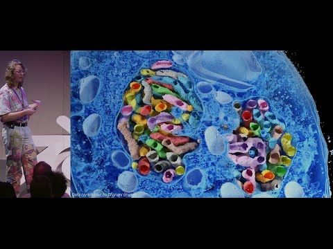
Microscopy Nodes: handling large biological volumes — Blender Conference 2025
YouTube video by Blender
youtu.be
September 19, 2025 at 7:35 AM
I gave a talk at Blender Conference yesterday, showing how biological volumes work, and how to visualize them in Blender 😋 youtu.be/WPajSWX730o?... #bcon25
Reposted by Kevin Terretaz
Still posting cytoskeleton videos, it seems. Actin this time.
Sample: Lifeact-eGFP in HeLa cells.
Modality: Airyscan confocal
Timestamp is mm:ss and the scale bar is 5 µm.
Sample: Lifeact-eGFP in HeLa cells.
Modality: Airyscan confocal
Timestamp is mm:ss and the scale bar is 5 µm.
November 23, 2025 at 3:21 AM
Still posting cytoskeleton videos, it seems. Actin this time.
Sample: Lifeact-eGFP in HeLa cells.
Modality: Airyscan confocal
Timestamp is mm:ss and the scale bar is 5 µm.
Sample: Lifeact-eGFP in HeLa cells.
Modality: Airyscan confocal
Timestamp is mm:ss and the scale bar is 5 µm.
Reposted by Kevin Terretaz

iPS cell-derived cardiac myocytes (heart muscle cells) typically beat about once per second, so I usually speed up the movies I post; otherwise, scrollers might miss the action. But every now and then, a cell looks like this in real time. #CellBiology
November 10, 2025 at 1:56 AM
iPS cell-derived cardiac myocytes (heart muscle cells) typically beat about once per second, so I usually speed up the movies I post; otherwise, scrollers might miss the action. But every now and then, a cell looks like this in real time. #CellBiology
Reposted by Kevin Terretaz
Attempt number seven at uploading this video of intermediate filaments in an enormous COS7 cell. I have a feeling the BlueSky compression will not do it any favors.
November 21, 2025 at 3:54 AM
Attempt number seven at uploading this video of intermediate filaments in an enormous COS7 cell. I have a feeling the BlueSky compression will not do it any favors.
Reposted by Kevin Terretaz

Happy #FluorescenceFriday. This beautiful video shows slice by slice section of a zebrafish embryo's head. The embryo is almost completely transparent, proving that zebrafish is an amazing model system for microscopy.
Brightfield, red = actin, blue = DAPI
📹: Postdoc Matyas BL (@Mongera lab, UCL)
Brightfield, red = actin, blue = DAPI
📹: Postdoc Matyas BL (@Mongera lab, UCL)
November 21, 2025 at 5:34 PM
Happy #FluorescenceFriday. This beautiful video shows slice by slice section of a zebrafish embryo's head. The embryo is almost completely transparent, proving that zebrafish is an amazing model system for microscopy.
Brightfield, red = actin, blue = DAPI
📹: Postdoc Matyas BL (@Mongera lab, UCL)
Brightfield, red = actin, blue = DAPI
📹: Postdoc Matyas BL (@Mongera lab, UCL)
Reposted by Kevin Terretaz

Finally, the first public release of #BigVolumeBrowser, so after teasers, you can try it yourself. For details, please check the announcement post (1/2)
forum.image.sc/t/bigvolumeb...
forum.image.sc/t/bigvolumeb...

BigVolumeBrowser: a new 3D multi volume/mesh/point clould (SMLM) data viewer
Hello everyone, I’d like to share with you another 3D viewer for FIJI, BigVolumeBrowser (full documentation link). It‘s a first initial public release, so there is still space for improvements. Le...
forum.image.sc
November 21, 2025 at 9:29 AM
Finally, the first public release of #BigVolumeBrowser, so after teasers, you can try it yourself. For details, please check the announcement post (1/2)
forum.image.sc/t/bigvolumeb...
forum.image.sc/t/bigvolumeb...
I updated the Image Viewer and the wiki page with many small bug fixes and features! If you haven't tried it yet, now it's even better ;)
#microscopy #ImageJ #FluorescenceFriday #microscopyMonday
#microscopy #ImageJ #FluorescenceFriday #microscopyMonday
My new ImageJ / Fiji toolkit is out 🔥! The goal is to make image handling & visualization easy, with an intuitive interface! Install it on Fiji with the "Image Viewer" update site
#microscopy #ImageJ #FluorescenceFriday #microscopyMonday
imagej.net/plugins/imag...
#microscopy #ImageJ #FluorescenceFriday #microscopyMonday
imagej.net/plugins/imag...
November 16, 2025 at 10:03 PM
I updated the Image Viewer and the wiki page with many small bug fixes and features! If you haven't tried it yet, now it's even better ;)
#microscopy #ImageJ #FluorescenceFriday #microscopyMonday
#microscopy #ImageJ #FluorescenceFriday #microscopyMonday
Reposted by Kevin Terretaz
This week's #FluorescenceFriday, we'll be treated with one of the classical models of #devbio, the neural crest cells. Here is a beautiful video of neural crest cells with GFP-tagged focal adhesion kinase (🔵) and LifeAct-RFP (🟣) migrating on a Fibronectin matrix.
📹: Adam Shellard
📹: Adam Shellard
November 14, 2025 at 5:45 PM
This week's #FluorescenceFriday, we'll be treated with one of the classical models of #devbio, the neural crest cells. Here is a beautiful video of neural crest cells with GFP-tagged focal adhesion kinase (🔵) and LifeAct-RFP (🟣) migrating on a Fibronectin matrix.
📹: Adam Shellard
📹: Adam Shellard
Reposted by Kevin Terretaz

I guess it never gets old...bc we love #CellMigration
ATP has many roles: energy currency, hydrotrope, and *danger signal*
Immune cells are faster after ATP exposure, F-actin (cyan) goes to the rear for nitro boost!
www.science.org/doi/full/10....
@focalplane.bsky.social #fluorescencefriday 🧪🔬
ATP has many roles: energy currency, hydrotrope, and *danger signal*
Immune cells are faster after ATP exposure, F-actin (cyan) goes to the rear for nitro boost!
www.science.org/doi/full/10....
@focalplane.bsky.social #fluorescencefriday 🧪🔬
November 14, 2025 at 8:38 PM
I guess it never gets old...bc we love #CellMigration
ATP has many roles: energy currency, hydrotrope, and *danger signal*
Immune cells are faster after ATP exposure, F-actin (cyan) goes to the rear for nitro boost!
www.science.org/doi/full/10....
@focalplane.bsky.social #fluorescencefriday 🧪🔬
ATP has many roles: energy currency, hydrotrope, and *danger signal*
Immune cells are faster after ATP exposure, F-actin (cyan) goes to the rear for nitro boost!
www.science.org/doi/full/10....
@focalplane.bsky.social #fluorescencefriday 🧪🔬
Reposted by Kevin Terretaz
🔬🍀
A timelapse movie (unpublished data) from the lab.
In the movie, the nucleus is moving towards the base of the polarized root hair cell.
A timelapse movie (unpublished data) from the lab.
In the movie, the nucleus is moving towards the base of the polarized root hair cell.
November 11, 2025 at 6:01 PM
🔬🍀
A timelapse movie (unpublished data) from the lab.
In the movie, the nucleus is moving towards the base of the polarized root hair cell.
A timelapse movie (unpublished data) from the lab.
In the movie, the nucleus is moving towards the base of the polarized root hair cell.
Reposted by Kevin Terretaz

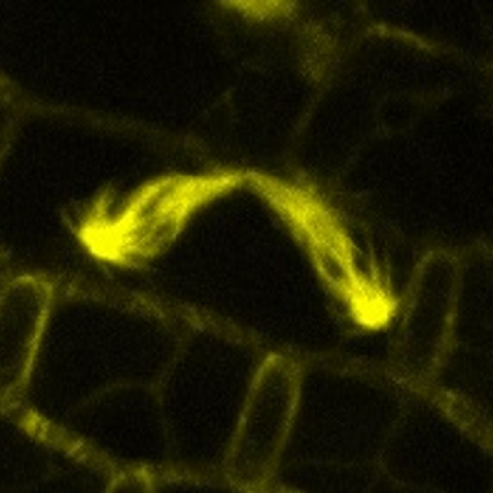
During my postdoc, I looked at hundreds of images like this 🤩
These are two apical caulonemal cells from moss Physcomitrium patens stained with MDY64 (shown in shades of orange). The natural autofluorescence of chlorophyll is in cyan.
#microscopymonday #moss #plantcells #plantmicroscopy
These are two apical caulonemal cells from moss Physcomitrium patens stained with MDY64 (shown in shades of orange). The natural autofluorescence of chlorophyll is in cyan.
#microscopymonday #moss #plantcells #plantmicroscopy

November 10, 2025 at 3:33 PM
During my postdoc, I looked at hundreds of images like this 🤩
These are two apical caulonemal cells from moss Physcomitrium patens stained with MDY64 (shown in shades of orange). The natural autofluorescence of chlorophyll is in cyan.
#microscopymonday #moss #plantcells #plantmicroscopy
These are two apical caulonemal cells from moss Physcomitrium patens stained with MDY64 (shown in shades of orange). The natural autofluorescence of chlorophyll is in cyan.
#microscopymonday #moss #plantcells #plantmicroscopy
Reposted by Kevin Terretaz

This image by MDI Bio Lab's Travis Carney is a #drosophila larval brain. Neural stem cells and neurons are marked, including axons that project into the brain. The flare in the center of each lobe is part of a learning and memory center in flies.
ZEISS Microscopy #microscopymonday 🧪 🤝
ZEISS Microscopy #microscopymonday 🧪 🤝

November 10, 2025 at 3:01 PM
This image by MDI Bio Lab's Travis Carney is a #drosophila larval brain. Neural stem cells and neurons are marked, including axons that project into the brain. The flare in the center of each lobe is part of a learning and memory center in flies.
ZEISS Microscopy #microscopymonday 🧪 🤝
ZEISS Microscopy #microscopymonday 🧪 🤝
Reposted by Kevin Terretaz

Happy #FluorescenceFriday from these primary astrocytes grown on glass slides and robotically stained on the Biocare Oncore Pro X! 🧠🔬🧪
Cyan = GFAP
Royal Purple = ALDH1L1
Salmon = Nuclear Counterstain
Cyan = GFAP
Royal Purple = ALDH1L1
Salmon = Nuclear Counterstain

November 7, 2025 at 7:26 PM
Happy #FluorescenceFriday from these primary astrocytes grown on glass slides and robotically stained on the Biocare Oncore Pro X! 🧠🔬🧪
Cyan = GFAP
Royal Purple = ALDH1L1
Salmon = Nuclear Counterstain
Cyan = GFAP
Royal Purple = ALDH1L1
Salmon = Nuclear Counterstain
Reposted by Kevin Terretaz
My new ImageJ / Fiji toolkit is out 🔥! The goal is to make image handling & visualization easy, with an intuitive interface! Install it on Fiji with the "Image Viewer" update site
#microscopy #ImageJ #FluorescenceFriday #microscopyMonday
imagej.net/plugins/imag...
#microscopy #ImageJ #FluorescenceFriday #microscopyMonday
imagej.net/plugins/imag...
November 4, 2025 at 10:40 PM
My new ImageJ / Fiji toolkit is out 🔥! The goal is to make image handling & visualization easy, with an intuitive interface! Install it on Fiji with the "Image Viewer" update site
#microscopy #ImageJ #FluorescenceFriday #microscopyMonday
imagej.net/plugins/imag...
#microscopy #ImageJ #FluorescenceFriday #microscopyMonday
imagej.net/plugins/imag...
Reposted by Kevin Terretaz

Two neurons went into a bar...

November 6, 2025 at 11:52 AM
Two neurons went into a bar...
Reposted by Kevin Terretaz
HL60 cells take on so many fun shapes as they migrate! This #InsightFromImaging data features cells prepared by Leanna and imaged on the @aicjanelia.bsky.social LLSM by @cmhobson.bsky.social
November 4, 2025 at 10:36 PM
HL60 cells take on so many fun shapes as they migrate! This #InsightFromImaging data features cells prepared by Leanna and imaged on the @aicjanelia.bsky.social LLSM by @cmhobson.bsky.social
My new ImageJ / Fiji toolkit is out 🔥! The goal is to make image handling & visualization easy, with an intuitive interface! Install it on Fiji with the "Image Viewer" update site
#microscopy #ImageJ #FluorescenceFriday #microscopyMonday
imagej.net/plugins/imag...
#microscopy #ImageJ #FluorescenceFriday #microscopyMonday
imagej.net/plugins/imag...
November 4, 2025 at 10:40 PM
My new ImageJ / Fiji toolkit is out 🔥! The goal is to make image handling & visualization easy, with an intuitive interface! Install it on Fiji with the "Image Viewer" update site
#microscopy #ImageJ #FluorescenceFriday #microscopyMonday
imagej.net/plugins/imag...
#microscopy #ImageJ #FluorescenceFriday #microscopyMonday
imagej.net/plugins/imag...
Reposted by Kevin Terretaz

Congratulations @centriolelab.bsky.social @dudinlab.bsky.social and @gautamdey.bsky.social for your publication in @cp-cell.bsky.social: Charting the landscape of #cytoskeletal diversity in #microbial eukaryotes #Expansion #Microscopy @sciencesunige.bsky.social
www.unige.ch/medias/en/20...
Congratulations @centriolelab.bsky.social @dudinlab.bsky.social and @gautamdey.bsky.social for your publication in @cp-cell.bsky.social: Charting the landscape of #cytoskeletal diversity in #microbial eukaryotes #Expansion #Microscopy @sciencesunige.bsky.social
www.unige.ch/medias/en/20...

November 3, 2025 at 1:34 PM
Congratulations @centriolelab.bsky.social @dudinlab.bsky.social and @gautamdey.bsky.social for your publication in @cp-cell.bsky.social: Charting the landscape of #cytoskeletal diversity in #microbial eukaryotes #Expansion #Microscopy @sciencesunige.bsky.social
www.unige.ch/medias/en/20...
Congratulations @centriolelab.bsky.social @dudinlab.bsky.social and @gautamdey.bsky.social for your publication in @cp-cell.bsky.social: Charting the landscape of #cytoskeletal diversity in #microbial eukaryotes #Expansion #Microscopy @sciencesunige.bsky.social
www.unige.ch/medias/en/20...
Reposted by Kevin Terretaz
In the spirit 👻 of Freaky Friday 🎃, here is a beautiful yet eery video of zebrafish retina development showing horizontal cells (🔵) finding their way out of the crowded amacrine (🟠) layer to settle beneath the photoreceptors. Happy #Halloween!
🎥: PhD student Rae Wong from the @nordenlab.bsky.social
🎥: PhD student Rae Wong from the @nordenlab.bsky.social
October 31, 2025 at 2:29 PM
In the spirit 👻 of Freaky Friday 🎃, here is a beautiful yet eery video of zebrafish retina development showing horizontal cells (🔵) finding their way out of the crowded amacrine (🟠) layer to settle beneath the photoreceptors. Happy #Halloween!
🎥: PhD student Rae Wong from the @nordenlab.bsky.social
🎥: PhD student Rae Wong from the @nordenlab.bsky.social
Reposted by Kevin Terretaz
An iPSC heart muscle cell forming sarcomeres videoed through a microscope by Emma Koory for 20 h before it dies most dramatically. This was our first try on a new microscope, and Emma now has the imaging parameters dialed in so her cells do not go POP! Alpha-actinin-2 is shown. #CellBiology
October 31, 2025 at 2:46 PM
An iPSC heart muscle cell forming sarcomeres videoed through a microscope by Emma Koory for 20 h before it dies most dramatically. This was our first try on a new microscope, and Emma now has the imaging parameters dialed in so her cells do not go POP! Alpha-actinin-2 is shown. #CellBiology