Alexander Krull
@alex-krull.bsky.social
100 followers
110 following
13 posts
Assistant Professor at Uni Birmingham. Working on Image processing, denoising, generative image models and other interesting things.
Posts
Media
Videos
Starter Packs
Alexander Krull
@alex-krull.bsky.social
· Aug 21
Reposted by Alexander Krull

Reposted by Alexander Krull


Reposted by Alexander Krull
Florian Jug
@florianjug.bsky.social
· Jul 14

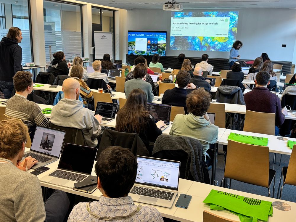
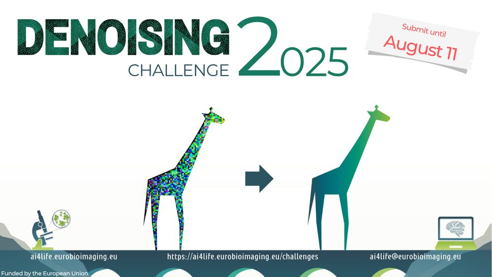
AI4Life
@ai4life.bsky.social
· Jun 27
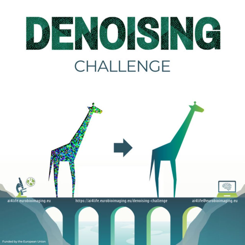
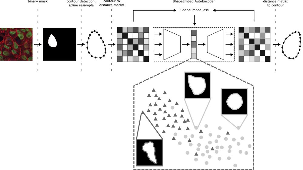
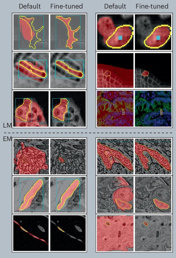
AI4Life Microscopy Denoising Challenge - Grand Challenge
Wellcome to AI4Life-MDC24! In this challenge, we want to focus on an unsupervised denoising of microscopy images. By participating, researchers can contribute to a critical area of scientific research...
ai4life-mdc24.grand-challenge.org
Reposted by Alexander Krull


Reposted by Alexander Krull
Reposted by Alexander Krull

Reposted by Alexander Krull
Reposted by Alexander Krull

Reposted by Alexander Krull

Alexander Krull
@alex-krull.bsky.social
· Mar 13

Reposted by Alexander Krull

Reposted by Alexander Krull

Reposted by Alexander Krull


Reposted by Alexander Krull


Alexander Krull
@alex-krull.bsky.social
· Feb 25
Reposted by Alexander Krull
Florian Jug
@florianjug.bsky.social
· Feb 17
Reposted by Alexander Krull