Cells & Development
@cellsdev.bsky.social
1.1K followers 430 following 31 posts
'Cells & Development' journal (formerly 'Mechanisms of Development'). Official journal of the International Society of Developmental Biology isdb.bsky.social
Posts Media Videos Starter Packs
Pinned
cellsdev.bsky.social
From January 2021 MoD will become “CELLS & DEVELOPMENT”, subtitle: “CELL AND DEVELOPMENTAL BIOLOGY AND THEIR QUANTITATIVE APPROCHES”. We expect that C&D will become a forum for interdisciplinary research combining biology, physics and mathematics to study cell and dev biology.
cellsdev.bsky.social
We need to be better! 😤
Reposted by Cells & Development
cellsdev.bsky.social
We often talk about what signals does the organiser release. But have you ever stopped and asked what does it mean to be the organiser? What cells make up the organiser? Where do they go? What do they do?
Well this review by Claudio Stern may give you the answers:
doi.org/10.1016/j.cd...
Cell movements around the organizer/node after stage HH4. Fate maps of the ectodermal (dorsal) layer of the organizer/node, based on Selleck and Stern (1991) (Selleck and Stern, 1991). Cell movements of populations contributing to the organizer. Stages of early chick development referred to in the text. The diagrams show some of the features used to classify the stages in relation to primitive streak formation.
Reposted by Cells & Development
cellsdev.bsky.social
In this intriguing paper published from the lab of @amartinezarias.bsky.social , they looked at the effects of size on morphology, tissue composition, and gene expression of gastruloid development. They found that AP elongation dynamics is size-dependent! Check it out here:
doi.org/10.1016/j.cd...
Reposted by Cells & Development
llobrienlab.bsky.social
For this #FluorescenceFriday, a gorgeous image of an adult mouse kidney labeled with AQP2 and alpha SMA antibodies. AQP2 (green) marks the collecting duct and distal connecting segment while SMA marks the arterial tree. Courtesy of talented postdoc Sarah McLarnon.
Image of a fluorescently labeled adult mouse kidney showing AQP2 staining of collecting ducts and connecting segment in green and alpha SMA staining of the arterial tree in magenta. The collecting ducts look like squiggly branches.
cellsdev.bsky.social
In this intriguing paper published from the lab of @amartinezarias.bsky.social , they looked at the effects of size on morphology, tissue composition, and gene expression of gastruloid development. They found that AP elongation dynamics is size-dependent! Check it out here:
doi.org/10.1016/j.cd...
cellsdev.bsky.social
Epithelial-to-mesenchymal transition or EMT is an integral part of development in animals. In this beautifully illustrated review from the lab of Magali Suzanne, they describe and compare and contrast the cellular mechanism of EMT across the different model organisms.
doi.org/10.1016/j.cd...
Fig. 1. EMT dynamics in developmental models. Fig. 2. A hypothetical model of EMT dynamics.
cellsdev.bsky.social
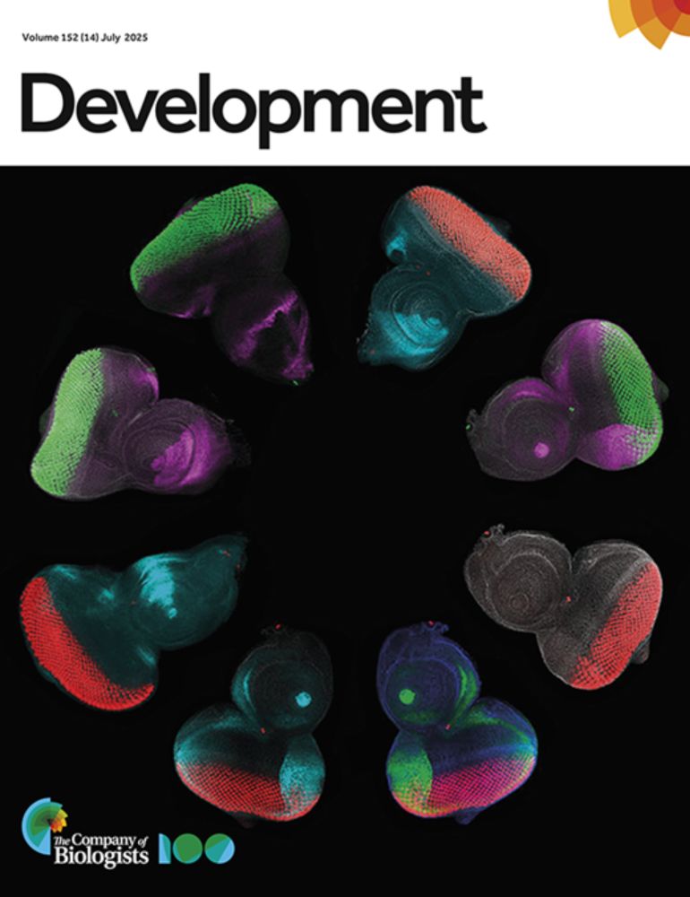
Our September issue cover is now online.
You can check out the issue here:
www.sciencedirect.com/journal/cell...
Reposted by Cells & Development
jcb.org
In 2019, Anna Jost & @jencwaters.bsky.social reviewed best practices for validating quantitative #microscopy methods & discuss strategies to avoid unconscious bias in imaging experiments rupress.org/jcb/article/...

📕 In #Reproducibility & Best Practices in Cell Biology: rupress.org/jcb/collecti...
Reposted by Cells & Development
v4sdb.bsky.social
We are grateful to all our sponsors who made the V4SDB Meeting 2025 in 🇸🇰 possible and accessible, specifically
Visegrád Fund, @biologists.bsky.social, @isdb.bsky.social, @cellsdev.bsky.social, @devdynamics.bsky.social, MUNI SCI, Allgene, BRIX, LABO-SK, and Merck Life Science.
Thank you!
Reposted by Cells & Development
mayorlab.bsky.social
Early Career Opportunity
Mentorship & career development, access to world-class facilities, and a collaborative, supportive environment. We are particularly interested in researchers in Synthetic Dev. Biol. Mechanobiology and AI . #AcademicJobs #EarlyCareerResearcher #UCL #DevelopmentalBiology
cellsdev.bsky.social
For decades, living embryos were the favourite tool to study development. However, their complexity hindered precise dissection of many processes. This comprehensive review provides a comparison between embryos and the power of gastruloids, opening up a new wave of dev bio.
doi.org/10.1016/j.cd...
Fig. 1. Schematic overview of mouse gastrulation Fig. 4. Different aspects of computational modelling of gastrulation. Fig. 3. Protein localisation, cell sorting behaviours and signalling pathway activation in gastruloids. Depicted are schematic representations based on integration of existing, fragmented data. Note that there is considerable variation in gastruloid development and existing data quality and resolution, and these cartoons represent our interpretation of average domains of immunofluorescence staining. Fig. 2. Schematic overview of signalling induced GRNs during gastrulation.
Reposted by Cells & Development
isdb.bsky.social
A cryosection from an embryo stained for the neural crest (🟠) using HCR, and DAPI (⚪).
Can you guess what embryo this is?
#FluorescenceFriday
Image credit: Ines Fernandez Mosquera, PhD student at @ucl.ac.uk
cellsdev.bsky.social
Many phenotypes from cells on plastic dishes just don't seem to translate to cells in vivo, particularly isolated primary cells. @raimonsunyer.bsky.social et al discussed how cells mechanically adapt, and the different ways we can mitigate this in this very helpful review:
doi.org/10.1016/j.cd...
The molecular clutch model offers insights into how cells adapt to rigid substrates such as tissue culture polystyrene (TCPS). Culturing cells on stiff substrates alters their mechanical phenotype. Stiff substrates, such as tissue culture polystyrene (TCPS), trigger adhesion reinforcement. Cell selection and mechanoadaptation when isolating primary cells. This figure illustrates how primary cells isolated from a tissue explant are affected by the stiffness of the culture surface. Strategies to mitigate stiff mechanoadaptation in cell culture.
Reposted by Cells & Development
cellsdev.bsky.social
What are the physics behind lumen formation? In this interesting and interdisciplinary review, Guillaume Salbreux et al described the physical laws from osmotic pressure to electrical potential that govern this biological process. Check it out here:
doi.org/10.1016/j.cd...
Examples of lumen formation. Mechanisms of lumen formation. Interactions of several lumens.
Reposted by Cells & Development
cellsdev.bsky.social
For something as classic as the Spemann-Mangold organizer first discovered in amphibians, the existence of such an organiser in mammals remains elusive. In this thought provoking review by Kate Mcdole et al, they discuss the evidence for and against such organiser in mouse.
doi.org/10.1016/j.cd...
Gastrula organizer in the mouse embryo. Cell migration and morphogenesis in the organizer. The AVE and PrCP in the formation of the head in mouse.
Reposted by Cells & Development
isdb.bsky.social
A classical neural crest migration. Even if the movie is a little bit shaky, their migration is still somehow mesmerising. It must be the stripes! 🤔 #fluorescencefriday

Movie by Kai Weissenbruch
Reposted by Cells & Development
anaisbailles.bsky.social
Very happy that the first article from my postdoc work in the Tomancak lab is now published @PNAS! www.pnas.org/doi/10.1073/.... We studied the self-organization of actin in aggregates made from Hydra cells. Thread below (1/9)
cellsdev.bsky.social
Can someone please add some beats 🎵🎵 to this video, we beg!
anaisbailles.bsky.social
What about stretch? We constrained the aggregates in agarose except for a hole. The tissue protrudes inside the hole, and the resulting stretch has a fast effect on actin alignment in the whole aggregate. Importantly, Wnt head organizers were not yet present in the protrusion. (6/9)
Reposted by Cells & Development
isdb.bsky.social
This is so cool! A good way for early career scientists to put your name out there and network too! Beautiful images are always eye catching. Register and we would love to see them too at ISDB!
socdevbio.bsky.social
Got stunning fluorescence microscopy images? 🌈🔬 SDB wants to feature YOUR work for #FluorescenceFriday! We highlight images from SDB members with a short description and the image creators 📸. Fill out the form to be featured: docs.google.com/forms/d/e/1F...