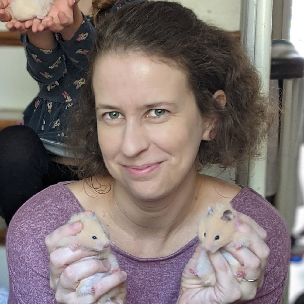
Emily Kolenbrander Ho
@emkolen.bsky.social
210 followers
430 following
23 posts
New asst prof at Claremont McKenna College | postdoc at Princeton, PhD from Stanford | devbio, signaling, and undergrad education
Posts
Media
Videos
Starter Packs
Pinned

Reposted by Emily Kolenbrander Ho
Reposted by Emily Kolenbrander Ho
Susanna Brantley
@drsusanna.bsky.social
· Jun 10
Reposted by Emily Kolenbrander Ho

Reposted by Emily Kolenbrander Ho
Reposted by Emily Kolenbrander Ho

Katherine W. Rogers
@katwrog.bsky.social
· Apr 21

An optogenetic toolkit for robust activation of FGF, BMP, and Nodal signaling in zebrafish
Cell signaling regulates a wide range of biological processes including development, homeostasis, and disease. Accessible technologies to precisely manipulate signaling have important applications in ...
www.biorxiv.org
Reposted by Emily Kolenbrander Ho

Mary Mirvis
@marymirvis.bsky.social
· Apr 19

A scoping study of the whole-cell imaging literature: a foundational corpus, potential for mesoscale data synthesis, and implications for standardization of an emerging field
The level of cellular organization bridging the mesoscale and whole-cell scale is coming into focus as a new frontier in cell biology. Great progress has been made in unraveling the complex physical a...
www.biorxiv.org
Reposted by Emily Kolenbrander Ho
jared toettcher
@toettch.bsky.social
· Mar 25
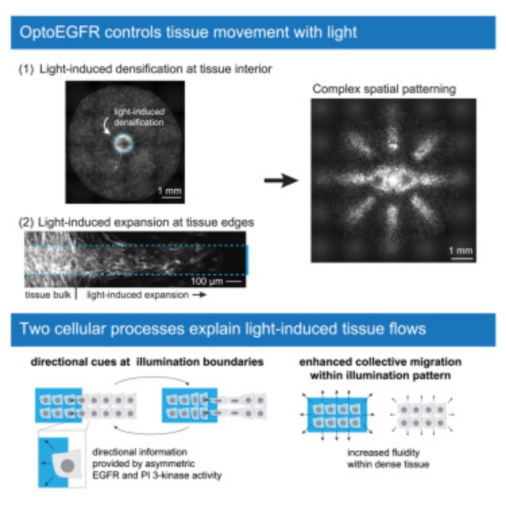
Large-scale control over collective cell migration using light-activated epidermal growth factor receptors
Programmable control over tissue movement is a fundamental challenge for tissue engineering
and wound healing. Suh, Thornton, et al. discovered that a light-controlled EGF receptor
controls long-range...
www.cell.com
Reposted by Emily Kolenbrander Ho
Eszter Posfai
@eposfai.bsky.social
· Mar 22
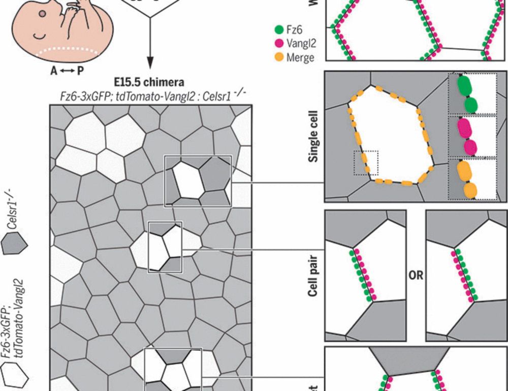
Epithelial polarization by the planar cell polarity complex is exclusively non–cell autonomous
For cells to polarize collectively along a tissue plane, asymmetrically localized planar cell polarity (PCP) complexes must form intercellular contacts between neighboring cells. Yet, it is unknown wh...
www.science.org
Reposted by Emily Kolenbrander Ho


Reposted by Emily Kolenbrander Ho
Reposted by Emily Kolenbrander Ho