
Leonhard Möckl
@lmoeckl.bsky.social
240 followers
230 following
36 posts
Professor of Nanooptical Imaging at FAU Erlangen-Nuremberg/CITABLE, Associated Group Leader at the MPI for the Science of Light. Glycobiology, optics, philosophy, and music.
Posts
Media
Videos
Starter Packs


Leonhard Möckl
@lmoeckl.bsky.social
· Sep 1
Reposted by Leonhard Möckl


Reposted by Leonhard Möckl
Marius Trollmann
@m-a-r-i-u-s.bsky.social
· Aug 28
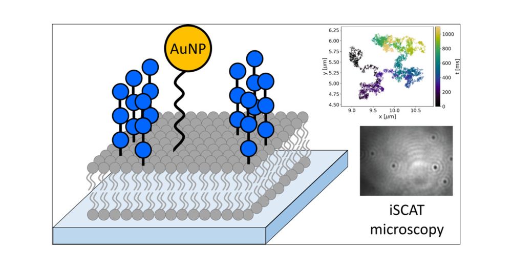
Bottom-up Investigation of Spatiotemporal Glycocalyx Dynamics with Interferometric Scattering Microscopy
Over recent decades, the glycocalyx, an extracellular organelle composed of a multitude of glycolipids, glycoproteins, proteoglycans, and glycoRNA, has gained considerable interest in cellular biology. While research in this field has revealed its tremendous importance in ever more aspects of physiological and pathological cellular processes, many of the principles that govern the role of the glycocalyx in these processes on a molecular level are still unknown. In order to unravel the fundamental laws underlying glycocalyx function, new technologies are required that enable the distinction between individual subprocesses within the intricate environment of the glycocalyx. Here, we establish an experimental platform to investigate the dynamics of the glycocalyx at the nanometer and microsecond length and time scales in a bottom-up fashion. We synthesized defined oligosaccharides and installed them on supported lipid bilayers. This way, synthetic glycolipids were assembled to glycocalyx model systems with tunable properties. By investigating these tunable model systems with interferometric scattering (iSCAT) microscopy, we gain access to the required spatiotemporal resolution. We found a strong correlation between the molecular structure of several investigated model glycans and global dynamics of the system. Our findings are corroborated by atomistic molecular dynamics simulations and coarse-grained Brownian dynamics simulations. Our results provide the first direct experimental evidence on the relationship between glycan structure, organization, and dynamics, offering a robust and versatile basis for a quantitative understanding of glycocalyx biology and physics at the molecular level.
pubs.acs.org
Leonhard Möckl
@lmoeckl.bsky.social
· Aug 28

Bottom-up Investigation of Spatiotemporal Glycocalyx Dynamics with Interferometric Scattering Microscopy
Over recent decades, the glycocalyx, an extracellular organelle composed of a multitude of glycolipids, glycoproteins, proteoglycans, and glycoRNA, has gained considerable interest in cellular biology...
pubs.acs.org
Reposted by Leonhard Möckl


Leonhard Möckl
@lmoeckl.bsky.social
· Jul 29
Leonhard Möckl
@lmoeckl.bsky.social
· Jul 28

Nature Nanotechnology
@natnano.nature.com
· Jul 28
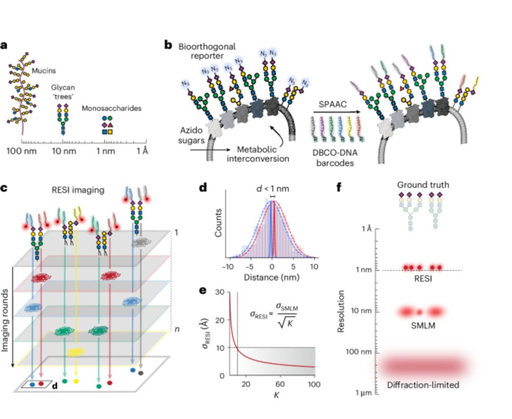
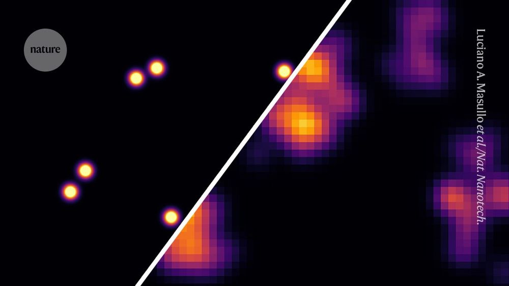
Ångström-resolution imaging of cell-surface glycans - Nature Nanotechnology
By combining bioorthogonal metabolic labelling and resolution enhancement through sequential imaging of DNA barcodes, the molecular organization of individual sugars in the native glycocalyx has been ...
www.nature.com


Leonhard Möckl
@lmoeckl.bsky.social
· May 2
Leonhard Möckl
@lmoeckl.bsky.social
· May 2


