Shashank Shekhar lab
@sshekhr.bsky.social
2.3K followers 3.1K following 85 posts
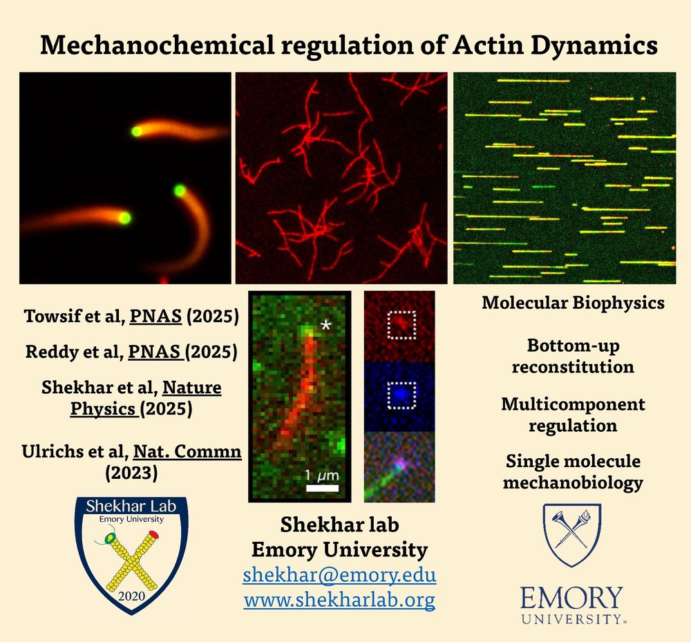
Prof. Shashank Shekhar @EmoryUniversity, Atlanta, USA Biophysicist interested in Actin dynamics and ciliary flows. Departments of Physics, Biochemistry and Cell Biology. Lab website : www.shekharlab.org
Posts Media Videos Starter Packs
Pinned
sshekhr.bsky.social
I am excited and honored to be named 'Cell Scientist to Watch' by Journal of Cell Science @biologists.bsky.social
Grateful to my team and mentors for their support.

Interested in working on actin? Hiring Postdocs/Grad students!
Reposted by Shashank Shekhar lab
sarawickstrom.bsky.social
My lab @mpi-muenster.bsky.social is looking for a computational biologist with a passion for imaging and spatial biology. A staff scientist position with long-term perspective! Apply and spread the word 🙏 jobs.mpi-muenster.mpg.de/jobposting/0...
Computational image analyst (m/f/d)
The Max Planck Institute for Molecular Biomedicine in Münster, Germany, has an opening for a
jobs.mpi-muenster.mpg.de
Reposted by Shashank Shekhar lab
syncellbiolab.bsky.social
How do cells keep their #actin “highways” stable?🚦
Our paper @PLOS Genetics reveals surprising teamwork (and backup plans!) between tropomyosin isoforms in yeast. #imaging #cytoskeleton #celldivision
This work was brilliantly led by @anubhavdhar.bsky.social @bagyasree.bsky.social
t.co/RvUrzad5tL
Reposted by Shashank Shekhar lab
daslabpombe.com
Our latest preprint on the role of the Arp2/3 complex in mitotic entry is now available @biorxiv. This work was done by Dhanya Kalathil, a very talented postdoc in my lab. The story started in 2019 when we were investigating the role of branched actin in cytokinesis. 1/
Reposted by Shashank Shekhar lab
stefandiezlab.bsky.social
Just started our @bcube-tud.bsky.social - organized Engineering Life 2025 #25EngLife conference in #Dresden! Excited to have everybody around!
sshekhr.bsky.social
📢📢Hiring 2 Research Assistants & 2 Postdocs at Emory University in Atlanta to study cytoskeletal biophysics/biochemistry. Please RT.

RAs: great for recent bachelor's/master's in Physics/Bio/Chem/Biochem. Email CV and interests to [email protected].

More info: www.shekharlab.org
Reposted by Shashank Shekhar lab
peiferlabunc.bsky.social
Last week I was privileged to join the Board of the Company of Biologists @biologists.bsky.social . They publish five journals across diverse fields of biology, including two of my favorites, @jcellsci.bsky.social & @dev-journal.bsky.social. 1/n
www.biologists.com
Description of journals published from the weblink
Reposted by Shashank Shekhar lab
epimechfc.bsky.social
Hi I’m @yamini-ravichan.bsky.social , a postdoc interested in the mechanical induction of topological defect-mediated morphogenesis. I use different organisms to study the role of topological defects shaping development & regeneration. Below is an insight to the field!
sshekhr.bsky.social
Presenting our lab's first PhD graduate, Dr. Heidi Ulrichs! 🎉🎓

Congratulations Heidi on such a successful PhD!
sshekhr.bsky.social
Actin ❤️
mag2art.bsky.social
The leading edge of a crawling cell videoed through a microscope. Watch the actin cytoskeleton in motion as it drives membrane protrusion and cell movement. It’s like watching the cell think with its feet. #CellBiology
sshekhr.bsky.social
That's a fantastic analogy! You will find the PDF on this link : www.shekharlab.org/_files/ugd/d...
www.shekharlab.org
Reposted by Shashank Shekhar lab
vikramalva.bsky.social
Excited to share our new preprint on HLp—a bacterial histone from Leptospira perolatii that forms stable tetramers and wraps ~60 bp of DNA: "DNA Wrapping by a Tetrameric Bacterial Histone" www.biorxiv.org/content/10.1...

@mpi-bio-fml.bsky.social
A snapshot of the HLp tetramer (purple) wrapping ~60 bp of DNA (green).
sshekhr.bsky.social
🎉🎉We’re on the cover of Nature Physics! @natphys.nature.com

Here is the original paper www.nature.com/articles/s41...
Reposted by Shashank Shekhar lab
ebrahim-lab.bsky.social
🧫 A gentle reminder that your gut is an actual work of art.
#FluorescenceFriday #BeneathTheScope
sshekhr.bsky.social
Thank you Tim!! I hope you are doing well as well!
Reposted by Shashank Shekhar lab
stcmicrobeblog.bsky.social
even if this 🧵 has already made its way to #ProtistsOnSky, it's a joy to repeat – so beautiful "rosettes" *)
...and the biophysics: stunning! 👏
sshekhr.bsky.social
📢Excited to share our new paper in Nature Physics
@naturephysics.bsky.social: Cooperative hydrodynamics accompany multicellular-like colonial organization in the unicellular Stentor!

How do single-celled organisms benefit from teamwork? Let’s dive in! #Multicellularity nature.com/articles/s41...?
sshekhr.bsky.social
Actin ❤️
christlet.bsky.social
Our PhD student Louis Romette is optimizing long-term live-cell imaging. He just casually dropped this 65-hour movie of a growing neuron (div 3 to 6) with knocked-in actin 🔥🕶️ #realtimemicroscopy
sshekhr.bsky.social
Thrilled to be featured in The New York Times! @nytimes.com

Fantastic piece by @jack-tamisiea.bsky.social on our discovery of the role of physical forces in evolution! With insightful comments from @wcratcliff.bsky.social!
sshekhr.bsky.social
When I think of the story about how you found them, I often imagine Jack and Sean coming out of the water like mermaids (or merman to be precise!) when you found them! 😁
Reposted by Shashank Shekhar lab
wallaceucsf.bsky.social
for a little back-story on how this project started in the Physiology course at the @mblscience.bsky.social , including how two of the authors become involved in the project after emerging from the water at "garbage beach" carrying lasers, check this out: wayback.archive-it.org/12248/201508...
Reposted by Shashank Shekhar lab
stcmicrobeblog.bsky.social
wonderful! great clip 👏
had never before seen 𝘚𝘵𝘦𝘯𝘵𝘰𝘳 competing with 𝘊𝘢𝘶𝘭𝘰𝘣𝘢𝘤𝘵𝘦𝘳 for rosette formation. no clear winner 🤩
#ProtistsOnSky #MicroSky
fluo­rescent image of rosettes of Caulobacter crescentus cells. Chromosomal DNA is labeled in blue (DAPI), cell membranes are in red (FM4-64). The fluorescently labeled protein is DivJ, a histidine kinase that localized at the base of the stalk (yellow). By Jennifer Heinritz, Jacobs-Wagner lab, Yale MSI. Cropped from https://www.instagram.com/p/BlBldEjBLzB/