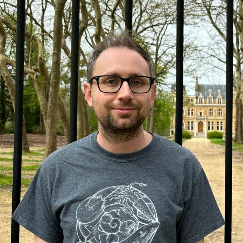
Ebrahim Lab
@ebrahim-lab.bsky.social
110 followers 110 following 9 posts
Lab focused on molecular force-generators and -sensors in living tissues, and all things microscopy @UVA School of Medicine. https://med.virginia.edu/ebrahim-lab/
Posts Media Videos Starter Packs
Reposted by Ebrahim Lab
jomaalab.bsky.social
Check out our new preprint on the discovery of a molecular switch in NAC that mediates nascent chain sorting on the ribosome and prevents mitochondrial protein mistargeting by SRP. A great collaboration with the Shan Lab @Caltech and the Qi Lab @UVA: www.biorxiv.org/content/10.1...
ebrahim-lab.bsky.social
Having seen your bingo card, we had to make it happen 😉!

Thank you for highlighting our work @hankgreen.bsky.social ! 🙏🏽
hankgreen.bsky.social
THIS WAS LITERALLY ON MY BINGO CARD!!
Scientists discover unknown organelle inside our cells
ebrahim-lab.bsky.social
Thank you, Ilya! :)
ebrahim-lab.bsky.social
Proposed mechanism of MVB formation
ebrahim-lab.bsky.social
Early for #MicroscopyMonday: #Hemifusomes- Newly discovered ESCRT-independent intermediates of intraluminal vesicle and multivesicular body formation! In collaboration with the Kachar Lab at the NIH and out today in @natcomms.nature.com : rdcu.be/emtQu

#cryoET #trafficking #organelle #hemifusion
ebrahim-lab.bsky.social
"Stereocilia"- actin-based skyscrapers that adorn sensory cells of our inner ear and covert vibrations into electrical signals—the first step in hearing and balance. #FluorescenceFriday
Reposted by Ebrahim Lab
sshekhr.bsky.social
🎉🎉We’re on the cover of Nature Physics! @natphys.nature.com

Here is the original paper www.nature.com/articles/s41...
ebrahim-lab.bsky.social
From stem cell to structure-
This human colonoid shows the self-organization of intestinal epithelial cells in 3D—nuclei in magenta, actin in green. A living miniature gut for your #FluorescenceFriday.
Reposted by Ebrahim Lab
thibautbrunet.bsky.social
A dream come true: the first expansion microscopy images of C. flexa 🤩 Generated by Mylan & Uzuki who learned from the best (@hiralshah.bsky.social @gautamdey.bsky.social @dudinlab.bsky.social). We will learn so much from these!
Expansion microscopy micrograph of the choanoflagellate Choanoeca flexa, vaguely evocative of a cactus. Red = tubulin, green = actin, blue = DNA Same with yellow = lipids (BODIPY)
Reposted by Ebrahim Lab
peiferlabunc.bsky.social
Only 29 slots left out of 200--register now!
peiferlabunc.bsky.social
Cell and developmental biologists: as you start looking at conferences in 2025, consider joining us at the 2025 Cell Contact & Adhesion GRC. We've assembled a strong and diverse program of speakers at the cutting-edge of science in our field 1/n Please repost
www.grc.org/cell-contact...
Poster for the meeting including speaker list and location
ebrahim-lab.bsky.social
🧫 A gentle reminder that your gut is an actual work of art.
#FluorescenceFriday #BeneathTheScope
Reposted by Ebrahim Lab
septinlab.bsky.social
Two months left for FASEB Conference - Septins: Spatial regulators of cell biology
Register now:
events.faseb.org/event/septin...
Come find out what these filaments do at the interface of the actin and microtubule cytoskeletons and cell membranes. You'll be hooked for life.
Reposted by Ebrahim Lab
poratshliomlab.bsky.social
Is liver zonation conserved from mice to humans? And how does fibrosis affect zonation in patients? Now, these questions are answered using scDVP. Some collaborations are meant to be! @carolineweiss.bsky.social@frosenberger.bsky.social @mannlab.bsky.social@mlsb-borgwardt.bsky.social
mannlab.bsky.social
Introducing our single-cell DVP framework combining strategic cell selection with protein gradient mapping algorithms. Analyzing hepatocyte proteomes across 18 human livers, revealing cross-species differences, and mapping disruptions in liver fibrosis.
www.biorxiv.org/content/10.1...
Reposted by Ebrahim Lab
dominikrobak.bsky.social
#MicroscopyMonday | Colon-scoping turned coral reef

Was imaging the colon when I accidentally stumbled into this glowing patch of vascularized white adipose tissue.

🔵 mT/mG mouse (mT-only) → red membranes shown in cyan

Accidental imaging hits different.

#IntravitalImaging #LiveImaging
Reposted by Ebrahim Lab
sergiocruzleon.bsky.social
Excited to share our preprint on the molecular architecture of heterochromatin in human cells 🧬🔬w/ @jpkreysing.bsky.social, @johannesbetz.bsky.social,
@marinalusic.bsky.social, Turoňová lab, @hummerlab.bsky.social @becklab.bsky.social @mpibp.bsky.social

🔗 Preprint here tinyurl.com/3a74uanv
Reposted by Ebrahim Lab
dominikrobak.bsky.social
#3DThursday | Intestinal villi in all their folded glory

These self-renewing, nutrient-absorbing structures are beautiful and functional.

🔴 Phalloidin
🔵 DAPI
🟢 Clvd cas-3 (extrusion in action!)

Fixed tissue 3D imaging = big insight, tiny resolution.
#Microscopy #3DImaging #biophysics #intestine
ebrahim-lab.bsky.social
Have cutting-edge cell biology research to share? Submit to Molecular Biology of the Cell (MBoC)- dedicated to advancing discovery and fostering collaboration.

Proud to be part of the editorial team supporting groundbreaking science. molbiolcell.org

#CellBiology #MBoC #Research
Molecular Biology of the Cell (MBoC)
molbiolcell.org