


Arindam Ghosh, PhD
@arindam92.bsky.social
Incoming Research Group Leader, DKFZ NCT WERA #Biophysics #Imaging #Immunology #Receptors
Reposted by Arindam Ghosh, PhD
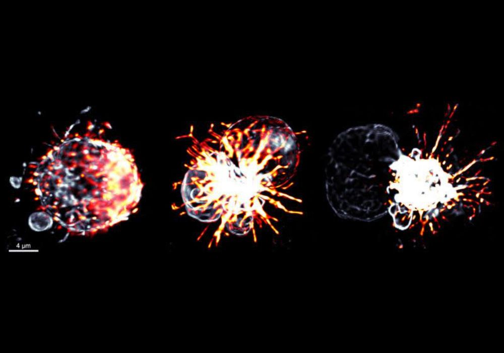
Got the last word in this great perspective on the state of fluorescent indicators in @nature.com by @dianakwon.bsky.social www.nature.com/articles/d41...

How genetically encoded sensors have lit up neuroscience
Tools that track specific molecules in neurons have enabled researchers to probe previously unexplored aspects of neurobiology — although important caveats remain.
www.nature.com
November 14, 2025 at 3:12 PM
Got the last word in this great perspective on the state of fluorescent indicators in @nature.com by @dianakwon.bsky.social www.nature.com/articles/d41...
Happy to share that I will be starting my independent research career as a Research Group Leader at the German Cancer Research Center (DKFZ). Starting from January 2026, my group will be based at DKFZ National Center for Tumor Diseases (NCT WERA), Würzburg, Germany.
November 11, 2025 at 1:33 PM
Happy to share that I will be starting my independent research career as a Research Group Leader at the German Cancer Research Center (DKFZ). Starting from January 2026, my group will be based at DKFZ National Center for Tumor Diseases (NCT WERA), Würzburg, Germany.
Reposted by Arindam Ghosh, PhD

👏 Congratulation to the 🏆 winners of Student Award held by #PicoQuant during 30th anniversary of our annual workshop on Single Molecule Spectroscopy and Ultra Sensitive Analysis in the Life Sciences. #WS30
We were proud to once again support the next generation of scientists. #PicoQuantStudentAward
We were proud to once again support the next generation of scientists. #PicoQuantStudentAward

September 26, 2025 at 11:12 AM
👏 Congratulation to the 🏆 winners of Student Award held by #PicoQuant during 30th anniversary of our annual workshop on Single Molecule Spectroscopy and Ultra Sensitive Analysis in the Life Sciences. #WS30
We were proud to once again support the next generation of scientists. #PicoQuantStudentAward
We were proud to once again support the next generation of scientists. #PicoQuantStudentAward
Reposted by Arindam Ghosh, PhD
🚨
New Preprint!
Ex-dSTORM resolves fine details of the molecular architecture of CCPs, the 8-nm periodicity of microtubules, and the docking site of synaptic vesicles at the presynapse of hippocampal neurons.
▶️ www.biorxiv.org/content/10.1...
New Preprint!
Ex-dSTORM resolves fine details of the molecular architecture of CCPs, the 8-nm periodicity of microtubules, and the docking site of synaptic vesicles at the presynapse of hippocampal neurons.
▶️ www.biorxiv.org/content/10.1...

August 19, 2025 at 3:32 PM
🚨
New Preprint!
Ex-dSTORM resolves fine details of the molecular architecture of CCPs, the 8-nm periodicity of microtubules, and the docking site of synaptic vesicles at the presynapse of hippocampal neurons.
▶️ www.biorxiv.org/content/10.1...
New Preprint!
Ex-dSTORM resolves fine details of the molecular architecture of CCPs, the 8-nm periodicity of microtubules, and the docking site of synaptic vesicles at the presynapse of hippocampal neurons.
▶️ www.biorxiv.org/content/10.1...
Reposted by Arindam Ghosh, PhD

Finally out in Nature Chem Bio:
SNAP-tag2 for faster and brighter protein labeling
www.nature.com/articles/s41...
Thank you Steffi and Veselin.
SNAP-tag2 for faster and brighter protein labeling
www.nature.com/articles/s41...
Thank you Steffi and Veselin.
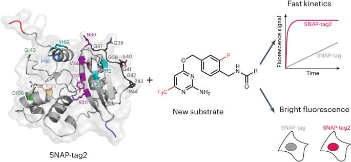
SNAP-tag2 for faster and brighter protein labeling - Nature Chemical Biology
SNAP-tag is a widespread tool for labeling protein for bioimaging. Now, Kühn et al. report SNAP-tag2 with increased labeling kinetics and brightness, which translates into a better performance in live...
www.nature.com
July 3, 2025 at 7:45 PM
Finally out in Nature Chem Bio:
SNAP-tag2 for faster and brighter protein labeling
www.nature.com/articles/s41...
Thank you Steffi and Veselin.
SNAP-tag2 for faster and brighter protein labeling
www.nature.com/articles/s41...
Thank you Steffi and Veselin.
Reposted by Arindam Ghosh, PhD
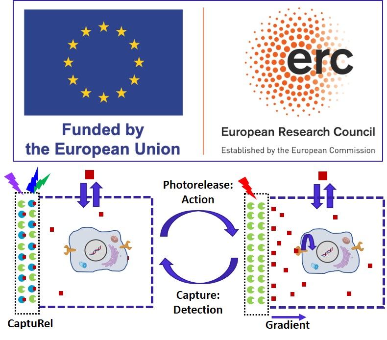
Great news: I received ERC Advanced grant #ERCAdG @erc.europa.eu CaptuRel! It aims at intelligent nanomaterials undergoing bioinspired cycle of capture and photorelease of bioactive molecules for sensing and controlling (bio)chemical gradients. Thanks to my Team, @cnrs.fr and @unistra.fr!
June 17, 2025 at 10:41 AM
Great news: I received ERC Advanced grant #ERCAdG @erc.europa.eu CaptuRel! It aims at intelligent nanomaterials undergoing bioinspired cycle of capture and photorelease of bioactive molecules for sensing and controlling (bio)chemical gradients. Thanks to my Team, @cnrs.fr and @unistra.fr!
Reposted by Arindam Ghosh, PhD
De novo designed bright, hyperstable rhodamine binders for fluorescence microscopy by Bo Huang and team: www.biorxiv.org/content/10.1...
June 26, 2025 at 1:50 PM
De novo designed bright, hyperstable rhodamine binders for fluorescence microscopy by Bo Huang and team: www.biorxiv.org/content/10.1...
Reposted by Arindam Ghosh, PhD

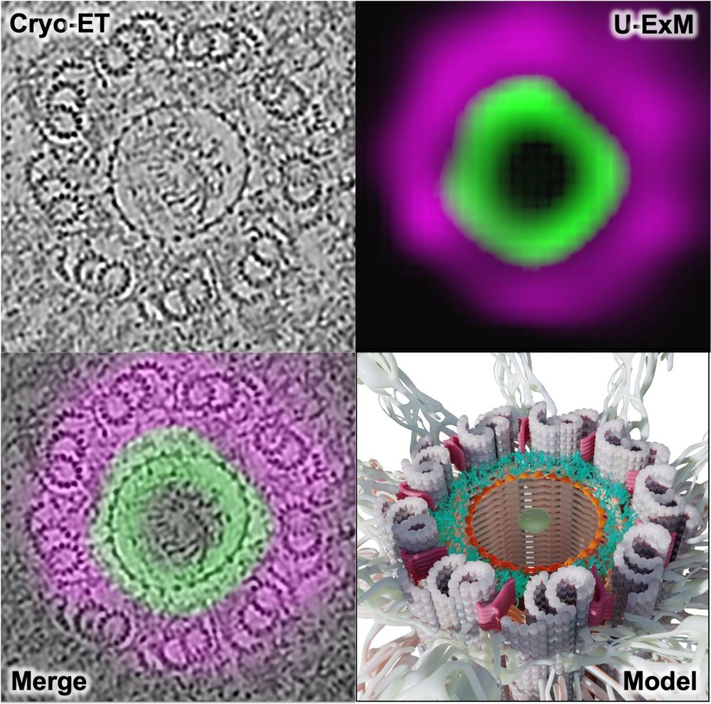
🚨 New preprint!
Using U-ExM + in situ cryo-ET, we show how C2CD3 builds an in-to-out radial architecture connecting the distal centriole lumen to its appendages. Great collab with @cellarchlab.com @chgenoud.bsky.social @stearnslab.bsky.social 🙌. #TeamTomo #UExM
www.biorxiv.org/content/10.1...
Using U-ExM + in situ cryo-ET, we show how C2CD3 builds an in-to-out radial architecture connecting the distal centriole lumen to its appendages. Great collab with @cellarchlab.com @chgenoud.bsky.social @stearnslab.bsky.social 🙌. #TeamTomo #UExM
www.biorxiv.org/content/10.1...
June 19, 2025 at 8:58 AM
🚨 New preprint!
Using U-ExM + in situ cryo-ET, we show how C2CD3 builds an in-to-out radial architecture connecting the distal centriole lumen to its appendages. Great collab with @cellarchlab.com @chgenoud.bsky.social @stearnslab.bsky.social 🙌. #TeamTomo #UExM
www.biorxiv.org/content/10.1...
Using U-ExM + in situ cryo-ET, we show how C2CD3 builds an in-to-out radial architecture connecting the distal centriole lumen to its appendages. Great collab with @cellarchlab.com @chgenoud.bsky.social @stearnslab.bsky.social 🙌. #TeamTomo #UExM
www.biorxiv.org/content/10.1...
Reposted by Arindam Ghosh, PhD
New paper out! Would you like to fluorescently label the plasma membrane in live, fixed or permeabilized cells? I tested different lipid structures and labelling approaches to decide what makes a good membrane labelling probe for such imaging experiments!
www.biorxiv.org/content/10.1...
www.biorxiv.org/content/10.1...

Plasma membrane labelling efficiency, internalization and partitioning of functionalized fluorescent lipids as a function of lipid structure
Labeling the plasma membrane for advanced imaging remains a significant challenge. For time-lapse live cell imaging, probe internalization and photobleaching are major limitations affecting most membr...
www.biorxiv.org
May 13, 2025 at 12:07 PM
New paper out! Would you like to fluorescently label the plasma membrane in live, fixed or permeabilized cells? I tested different lipid structures and labelling approaches to decide what makes a good membrane labelling probe for such imaging experiments!
www.biorxiv.org/content/10.1...
www.biorxiv.org/content/10.1...
Reposted by Arindam Ghosh, PhD
Very excited to present our latest work: SPINNA, an analysis framework and software package for single-protein resolution data! 🖥️🤩
We can directly quantify stoichiometry and oligomerization from super-res (DNA-PAINT, RESI) images!! 🧬🎨
We can directly quantify stoichiometry and oligomerization from super-res (DNA-PAINT, RESI) images!! 🧬🎨

Spatial and stoichiometric in situ analysis of biomolecular oligomerization at single-protein resolution
We are excited to present our latest work published in @natcomms.nature.com
www.nature.com/articles/s41...
We are excited to present our latest work published in @natcomms.nature.com
www.nature.com/articles/s41...
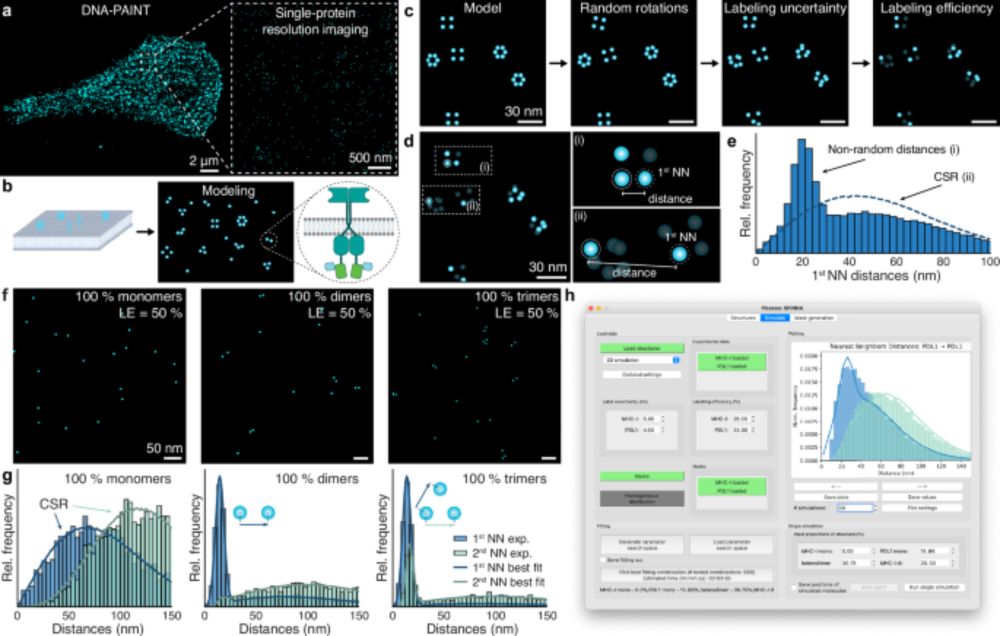
Spatial and stoichiometric in situ analysis of biomolecular oligomerization at single-protein resolution - Nature Communications
Extracting quantitative information on biomolecular oligomerisation with high resolution remains a significant challenge. Here, the authors propose SPINNA, a framework that compares nearest-neighbour ...
www.nature.com
May 7, 2025 at 2:56 PM
Very excited to present our latest work: SPINNA, an analysis framework and software package for single-protein resolution data! 🖥️🤩
We can directly quantify stoichiometry and oligomerization from super-res (DNA-PAINT, RESI) images!! 🧬🎨
We can directly quantify stoichiometry and oligomerization from super-res (DNA-PAINT, RESI) images!! 🧬🎨
Reposted by Arindam Ghosh, PhD
Collaborative work with the Shechtman Lab and @heilemannlab.bsky.social.
Check it out here: www.biorxiv.org/content/10.1...
Check it out here: www.biorxiv.org/content/10.1...

One-click image reconstruction in single-molecule localization microscopy via deep learning
Deep neural networks have led to significant advancements in microscopy image generation and analysis. In single-molecule localization based super-resolution microscopy, neural networks are capable of...
www.biorxiv.org
April 22, 2025 at 1:16 PM
Collaborative work with the Shechtman Lab and @heilemannlab.bsky.social.
Check it out here: www.biorxiv.org/content/10.1...
Check it out here: www.biorxiv.org/content/10.1...
Our work on lattice light-sheet TDI-DNA-PAINT and anti-CD20 immunotherapy antibodies is now featured in The Scientist magazine www.the-scientist.com. Many thanks to Sneha Khedkar for writing this fantastic piece. www.the-scientist.com/new-microsco...
New Microscopy Technique Challenges Therapeutic Antibody Classification
A super-resolution microscopy technique offers an unparalleled glimpse into how monoclonal antibodies bind to their targets on cancer cells to induce cell death.
www.the-scientist.com
March 25, 2025 at 1:01 PM
Our work on lattice light-sheet TDI-DNA-PAINT and anti-CD20 immunotherapy antibodies is now featured in The Scientist magazine www.the-scientist.com. Many thanks to Sneha Khedkar for writing this fantastic piece. www.the-scientist.com/new-microsco...
Reposted by Arindam Ghosh, PhD
🔬Happy to share our new preprint, where brightness is used to identify single molecule emission in 2D/3D, with a single camera ! Part of the PhD work of @laurent-le.bsky.social and Surabhi, great collaboration with @emmanuelfort.bsky.social from @instlangevin.bsky.social @ismolab.bsky.social #SMLM

March 4, 2025 at 8:58 AM
🔬Happy to share our new preprint, where brightness is used to identify single molecule emission in 2D/3D, with a single camera ! Part of the PhD work of @laurent-le.bsky.social and Surabhi, great collaboration with @emmanuelfort.bsky.social from @instlangevin.bsky.social @ismolab.bsky.social #SMLM
Reposted by Arindam Ghosh, PhD

Congratulations to Rice Chemistry Prof. Anna-Karin Gustavsson for receiving the NSF CAREER.
@gustavssonlab.bsky.social #NSFCAREER #RiceChemistry
@gustavssonlab.bsky.social #NSFCAREER #RiceChemistry
Anna-Karin Gustavsson on LinkedIn: Rice’s Gustavsson receives NSF CAREER Award to investigate dynamics of…
Very honored and grateful to receive the NSF CAREER Award! Big thanks to the National Science Foundation (NSF) and to my fantastic research team and…
www.linkedin.com
March 4, 2025 at 6:31 PM
Congratulations to Rice Chemistry Prof. Anna-Karin Gustavsson for receiving the NSF CAREER.
@gustavssonlab.bsky.social #NSFCAREER #RiceChemistry
@gustavssonlab.bsky.social #NSFCAREER #RiceChemistry
Reposted by Arindam Ghosh, PhD
Happy to share our newest paper in Ang. Chem. and big thanks to @nkaredla.bsky.social, @faldalf.bsky.social and @tao-chen.bsky.social 🙏 onlinelibrary.wiley.com/doi/10.1002/...

Leaflet‐specific Structure and Dynamics of Solid and Polymer Supported Lipid Bilayers
Polymer-supported or tethered lipid bilayers serve as versatile platforms for mimicking plasma membrane structure and dynamics, yet the impact of polymer supports on lipid bilayers remains largely un...
onlinelibrary.wiley.com
March 11, 2025 at 7:58 AM
Happy to share our newest paper in Ang. Chem. and big thanks to @nkaredla.bsky.social, @faldalf.bsky.social and @tao-chen.bsky.social 🙏 onlinelibrary.wiley.com/doi/10.1002/...
Reposted by Arindam Ghosh, PhD

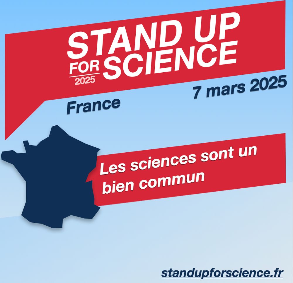
🧪Paris Jussieu right now #standupforscience

March 7, 2025 at 12:41 PM
🧪Paris Jussieu right now #standupforscience
Reposted by Arindam Ghosh, PhD

Science, research & public health face unprecedented attacks in the U.S.
We stand with our American colleagues & support the Stand Up for Science call.
Join the global march tomorrow, incl. in Paris! 🧪✊
📍 March 7, 13:30 – Place Jussieu
🔗 More info: standupforscience.fr
#StandUpForScience
We stand with our American colleagues & support the Stand Up for Science call.
Join the global march tomorrow, incl. in Paris! 🧪✊
📍 March 7, 13:30 – Place Jussieu
🔗 More info: standupforscience.fr
#StandUpForScience
March 6, 2025 at 2:24 PM
Science, research & public health face unprecedented attacks in the U.S.
We stand with our American colleagues & support the Stand Up for Science call.
Join the global march tomorrow, incl. in Paris! 🧪✊
📍 March 7, 13:30 – Place Jussieu
🔗 More info: standupforscience.fr
#StandUpForScience
We stand with our American colleagues & support the Stand Up for Science call.
Join the global march tomorrow, incl. in Paris! 🧪✊
📍 March 7, 13:30 – Place Jussieu
🔗 More info: standupforscience.fr
#StandUpForScience
Reposted by Arindam Ghosh, PhD
I'm thrilled to share that our review article, "Molecular Level Super-Resolution Fluorescence Imaging," has now been published online (www.annualreviews.org/content/jour...)! We provide a comprehensive overview of fluorescence super-res methods that push the limits towards molecular-scale imaging.
Molecular Level Super-Resolution Fluorescence Imaging | Annual Reviews
Over the last 30 years, fluorescence microscopy, renowned for its sensitivity and specificity, has undergone a revolution in resolving ever-smaller details. This advancement began with stimulated emis...
www.annualreviews.org
February 18, 2025 at 3:52 PM
I'm thrilled to share that our review article, "Molecular Level Super-Resolution Fluorescence Imaging," has now been published online (www.annualreviews.org/content/jour...)! We provide a comprehensive overview of fluorescence super-res methods that push the limits towards molecular-scale imaging.
Reposted by Arindam Ghosh, PhD

Online now! Fluorescent labeling strategies for molecular bioimaging. Marcel Streit, Made Budiarta, Marvin Jungblut, and Gerti Beliu. www.cell.com/biophysrepor...

Fluorescent labeling strategies for molecular bioimaging
Super-resolution microscopy (SRM) has transformed biological imaging by circumventing
the diffraction limit of light and enabling the visualization of cellular structures
and processes at the molecula...
www.cell.com
February 14, 2025 at 5:08 PM
Online now! Fluorescent labeling strategies for molecular bioimaging. Marcel Streit, Made Budiarta, Marvin Jungblut, and Gerti Beliu. www.cell.com/biophysrepor...
Reposted by Arindam Ghosh, PhD
🆕 Our new functionalized docetaxel probe enables superior visualization of dense mitotic spindle structures during cell division by expansion microscopy!
▶️ pubs.acs.org/doi/10.1021/...
▶️ pubs.acs.org/doi/10.1021/...
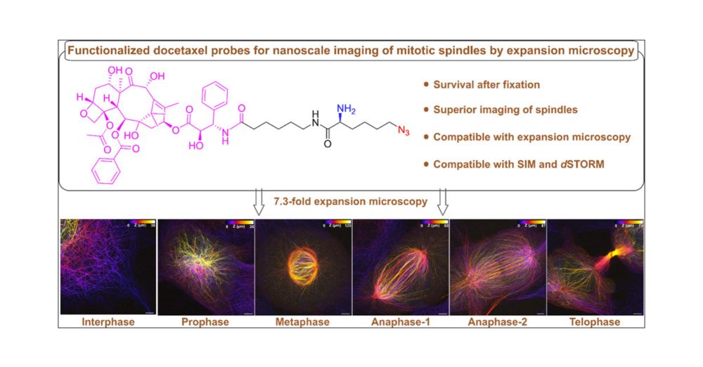
Functionalized Docetaxel Probes for Refined Visualization of Mitotic Spindles by Expansion Microscopy
Visualizing the ultrastructure of mitotic spindles, the macromolecular machines that segregate chromosomes during mitosis, by fluorescence imaging remains challenging. Here we introduce an azido- and ...
pubs.acs.org
February 12, 2025 at 12:49 PM
🆕 Our new functionalized docetaxel probe enables superior visualization of dense mitotic spindle structures during cell division by expansion microscopy!
▶️ pubs.acs.org/doi/10.1021/...
▶️ pubs.acs.org/doi/10.1021/...
Reposted by Arindam Ghosh, PhD
Check it out, powerful new method for optical imaging of the live ECM, from @hhmijanelia.bsky.social Group Leader Kayvon Pedram #proudofalumni #glycotime
www.nature.com/articles/s41...
www.nature.com/articles/s41...
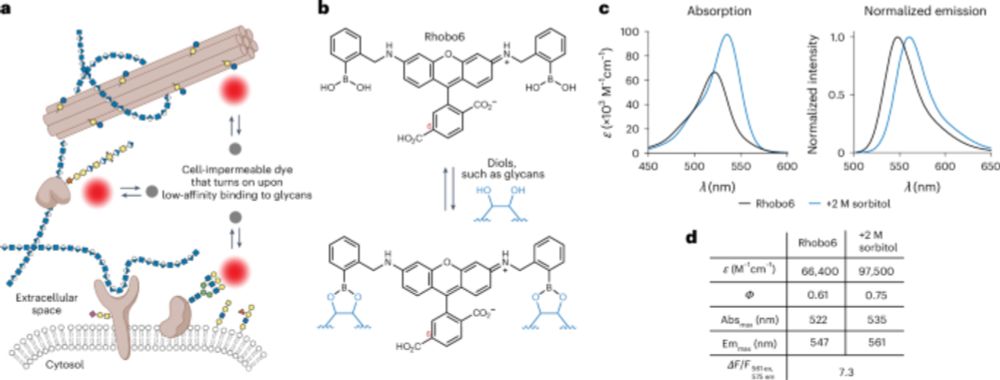
Live imaging of the extracellular matrix with a glycan-binding fluorophore - Nature Methods
Rhobo6 is a cell-impermeable small-molecule fluorophore that displays reversible fluorogenic binding to glycans, making it a general, wash-free, and non-perturbative label for the extracellular matrix...
www.nature.com
February 6, 2025 at 10:59 PM
Check it out, powerful new method for optical imaging of the live ECM, from @hhmijanelia.bsky.social Group Leader Kayvon Pedram #proudofalumni #glycotime
www.nature.com/articles/s41...
www.nature.com/articles/s41...
Reposted by Arindam Ghosh, PhD

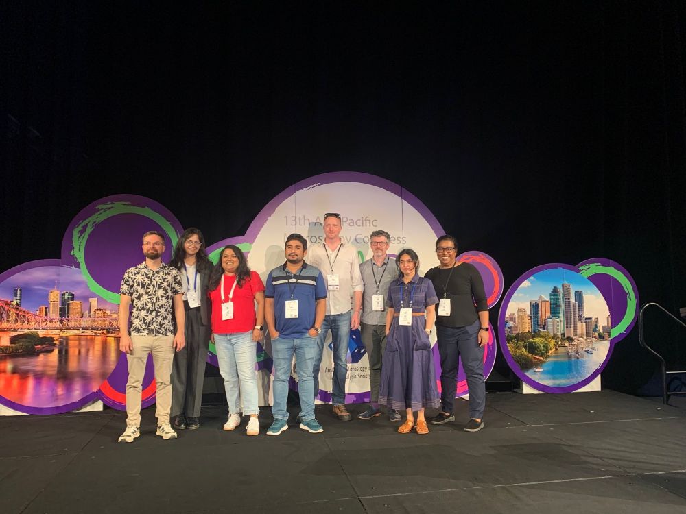
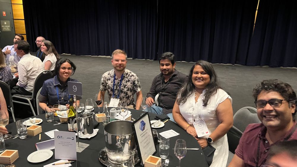
Aaaand that’s a wrap at Asia Pacific Microscopy Congress 2025 in sunny Brisbane, attended by a big team from Single Molecule Science. Lots of exciting new 🔬 tools and translation in biomedical investigations. @ijayas.bsky.social @arindam92.bsky.social
February 7, 2025 at 10:19 AM
Aaaand that’s a wrap at Asia Pacific Microscopy Congress 2025 in sunny Brisbane, attended by a big team from Single Molecule Science. Lots of exciting new 🔬 tools and translation in biomedical investigations. @ijayas.bsky.social @arindam92.bsky.social
Reposted by Arindam Ghosh, PhD
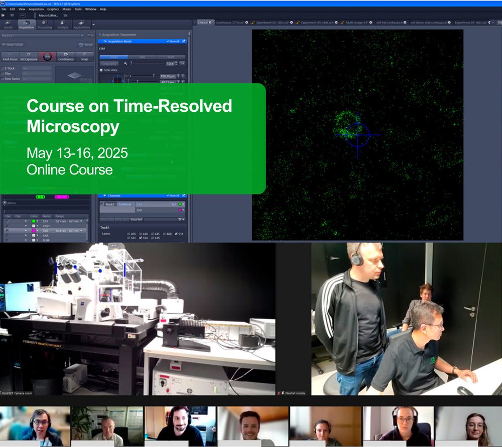
Want to explore the world of time-resolved fluorescence #microscopy? Sign up for our online course and benefit from theoretical as well as practical sessions in an interactive format. Topics: data analysis, FCS, FRET, FLIM, and more. May 13-16, 2025. ➡️ www.microscopy-course.org
February 4, 2025 at 10:52 AM
Want to explore the world of time-resolved fluorescence #microscopy? Sign up for our online course and benefit from theoretical as well as practical sessions in an interactive format. Topics: data analysis, FCS, FRET, FLIM, and more. May 13-16, 2025. ➡️ www.microscopy-course.org
Reposted by Arindam Ghosh, PhD

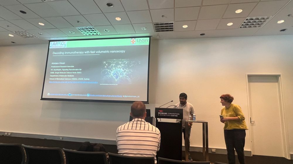
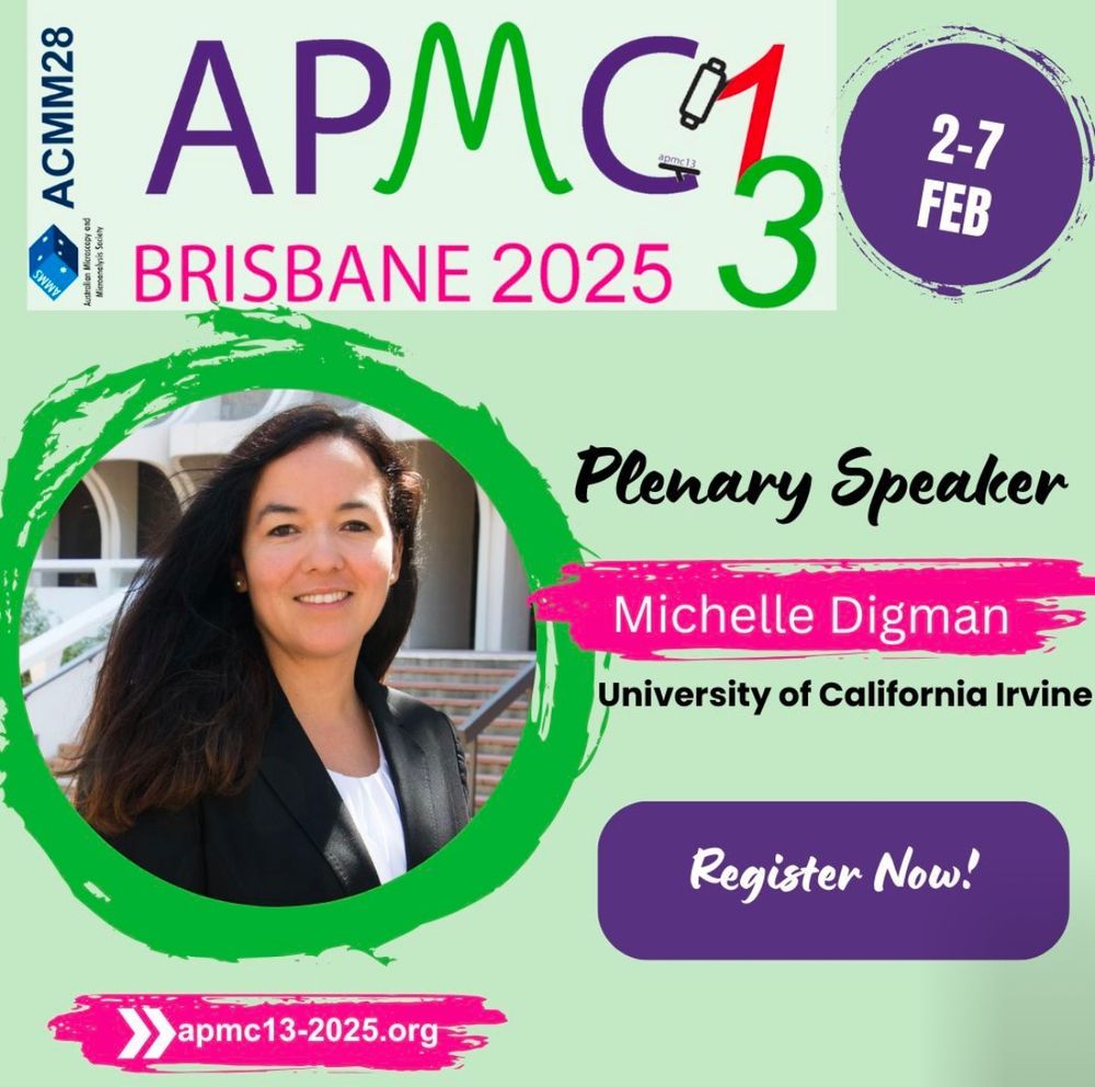
Thrilled and honored to present a plenary talk at the 13th Asia Pacific Microscopy Congress (APMC13) in Brisbane, Australia, on February 7, 2025, hosted by the Australian Microscopy and Microanalysis Society (AMMS).
February 2, 2025 at 7:16 PM
Thrilled and honored to present a plenary talk at the 13th Asia Pacific Microscopy Congress (APMC13) in Brisbane, Australia, on February 7, 2025, hosted by the Australian Microscopy and Microanalysis Society (AMMS).