


Vanja Tepavcevic
@tepavcevicvanja.bsky.social
Neurobiologist.Oligodendrocytes/myelin/remyelination/energy metabolism.
Pinned
Monocarboxylate transporter 2 is required for the maintenance of myelin and axonal integrity by oligodendrocytes https://www.biorxiv.org/content/10.1101/2025.01.10.632306v1
Interested in #oligodendrocyte metabolism and/or #multiplesclerosis? Our paper is now at Biorxiv! We show that oligos express MCT2, the high affinity monocarboxylate transporter, well known for supplying neurons with astrocyte-derived lactate (astro-neuron lactate shuttle)👇
Reposted by Vanja Tepavcevic

Poor heart health in middle age linked to dementia in old age – new study theconversation.com/poor-heart-h...
#Alzheimers #dementia #health #wellness
#Alzheimers #dementia #health #wellness

Poor heart health in middle age linked to dementia in old age – new study
A 25-year study reveals that silent heart damage in your 50s can predict dementia risk decades later.
theconversation.com
November 12, 2025 at 1:04 PM
Poor heart health in middle age linked to dementia in old age – new study theconversation.com/poor-heart-h...
#Alzheimers #dementia #health #wellness
#Alzheimers #dementia #health #wellness
Reposted by Vanja Tepavcevic

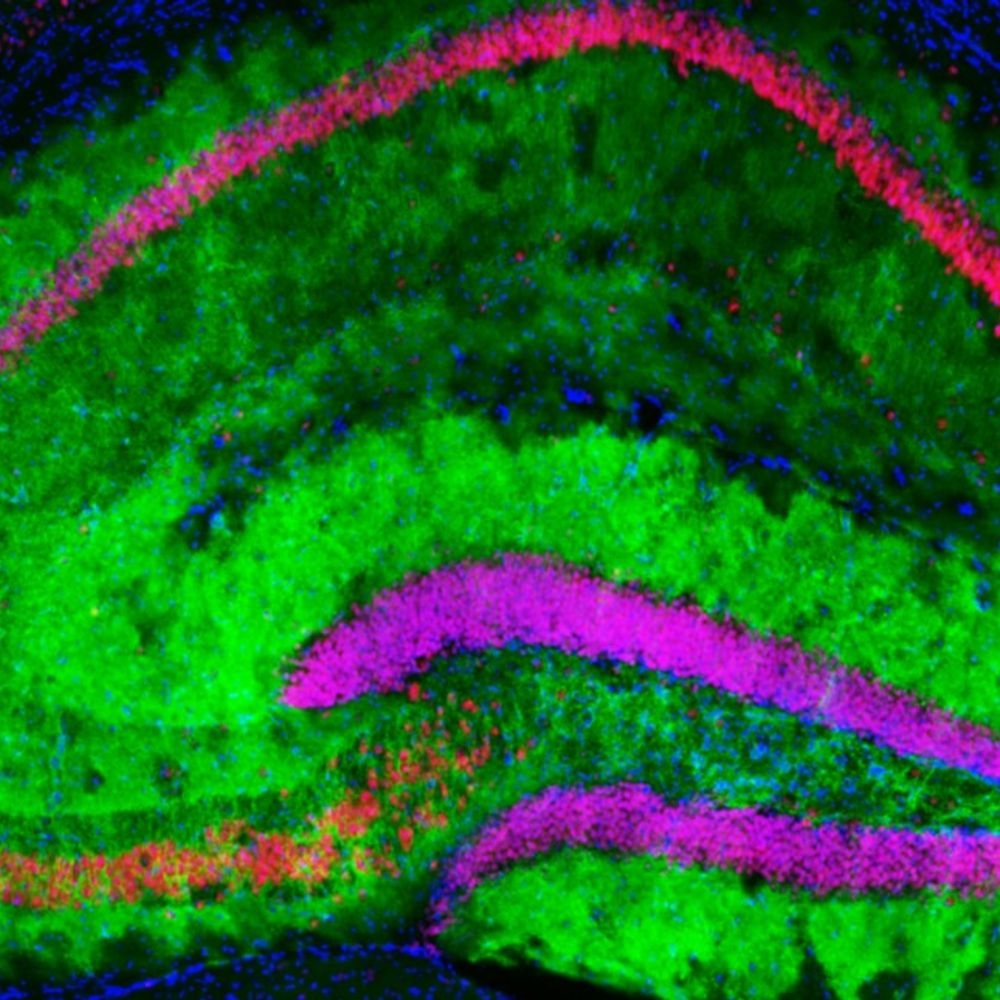
Looking forward to presenting our work on oligodendrocyte dysfunction in a rare neurodegenerative movement disorder X-linked Dystonia Parkinsonism. Come say hi if you're around on Tuesday, Nov 18 between 1-5 pm! @sfn.org #SfN2025 #SfN25
@sfn.org #SfN25 #SanDiego starts this Saturday. The lab is going to be there in full force - see our presentations below.
Represented this year by @borderbiologist.bsky.social, Nick Kathman, @mrodea.bsky.social, @melcooperphd.bsky.social, @pryprk.bsky.social, @kerrylimberg.bsky.social.
(1/2)
Represented this year by @borderbiologist.bsky.social, Nick Kathman, @mrodea.bsky.social, @melcooperphd.bsky.social, @pryprk.bsky.social, @kerrylimberg.bsky.social.
(1/2)

November 10, 2025 at 11:09 PM
Reposted by Vanja Tepavcevic

Local #mRNAtranslation in polarized cells is well studied in neurons but what about #microglia? This study shows that #inflammation enhances microglial #RNA localization & translation via RNA-binding protein IMP1/ZBP1, regulating morphology, motility & phagocytosis @plosbiology.org 🧪 plos.io/486ZLMS

November 11, 2025 at 9:48 AM
Local #mRNAtranslation in polarized cells is well studied in neurons but what about #microglia? This study shows that #inflammation enhances microglial #RNA localization & translation via RNA-binding protein IMP1/ZBP1, regulating morphology, motility & phagocytosis @plosbiology.org 🧪 plos.io/486ZLMS
Reposted by Vanja Tepavcevic
📢 Interested in #lipidomics and #dementia? Join us tomorrow 👇
🚨Join our next @DEMONNetworkUK #Genetics and #Omics joint with #Biomarkers meeting on THURSDAY 6th November at 2.30 PM (UK Time). Delighted to have Asger Wretlind as invited speaker – “Unsaturated #Lipid Deficits: A #Female Specific #Alzheimer's Signature”
Join the network or DM for more info!
Join the network or DM for more info!
November 5, 2025 at 9:47 PM
📢 Interested in #lipidomics and #dementia? Join us tomorrow 👇
Reposted by Vanja Tepavcevic

Últimos días para presentar candidaturas al Comité Científico Técnico de la @AgEInves.
📅 Plazo hasta el 10 de noviembre
ℹ️ Procedimiento y detalles: www.aei.gob.es/sobre-aei/or...
#Últimallamada #ComitéCientíficoTécnico #EvaluaciónCientífica
📅 Plazo hasta el 10 de noviembre
ℹ️ Procedimiento y detalles: www.aei.gob.es/sobre-aei/or...
#Últimallamada #ComitéCientíficoTécnico #EvaluaciónCientífica

November 5, 2025 at 10:50 AM
Últimos días para presentar candidaturas al Comité Científico Técnico de la @AgEInves.
📅 Plazo hasta el 10 de noviembre
ℹ️ Procedimiento y detalles: www.aei.gob.es/sobre-aei/or...
#Últimallamada #ComitéCientíficoTécnico #EvaluaciónCientífica
📅 Plazo hasta el 10 de noviembre
ℹ️ Procedimiento y detalles: www.aei.gob.es/sobre-aei/or...
#Últimallamada #ComitéCientíficoTécnico #EvaluaciónCientífica
Reposted by Vanja Tepavcevic
Join us at the last IBRO Connect of 2025 during #SfN2025. Connect with fellow #neuroscientists & join us in celebrating this year’s achievements in a fun atmosphere!
17 Nov | 19:00–21:00 PST at Venue 808, San Diego
Limited seats: https://ibro.org/event/ibro-connect-networking-event-in-san-diego/
17 Nov | 19:00–21:00 PST at Venue 808, San Diego
Limited seats: https://ibro.org/event/ibro-connect-networking-event-in-san-diego/

November 5, 2025 at 8:00 AM
Join us at the last IBRO Connect of 2025 during #SfN2025. Connect with fellow #neuroscientists & join us in celebrating this year’s achievements in a fun atmosphere!
17 Nov | 19:00–21:00 PST at Venue 808, San Diego
Limited seats: https://ibro.org/event/ibro-connect-networking-event-in-san-diego/
17 Nov | 19:00–21:00 PST at Venue 808, San Diego
Limited seats: https://ibro.org/event/ibro-connect-networking-event-in-san-diego/
Reposted by Vanja Tepavcevic
Thrilled to publish our new study on #mitochondrial ROS coming from complex III, revealing this key site in #astrocytes 🎯as a crucial immunometabolic signal transducer🤯 and potential therapeutic target for #dementia #FTD 💊 rdcu.be/eOc0m 👈💪 Great commentary by H. Pan and F. Yin🤩! tinyurl.com/4hdytw4c

November 4, 2025 at 6:30 PM
Thrilled to publish our new study on #mitochondrial ROS coming from complex III, revealing this key site in #astrocytes 🎯as a crucial immunometabolic signal transducer🤯 and potential therapeutic target for #dementia #FTD 💊 rdcu.be/eOc0m 👈💪 Great commentary by H. Pan and F. Yin🤩! tinyurl.com/4hdytw4c
Reposted by Vanja Tepavcevic
@biorxivpreprint.bsky.social Regulatory T-cells in multiple sclerosis produce IL-10 in the central nervous system but are activated by Epstein-Barr Virus
www.biorxiv.org/content/10.1...
www.biorxiv.org/content/10.1...

October 24, 2025 at 7:14 PM
@biorxivpreprint.bsky.social Regulatory T-cells in multiple sclerosis produce IL-10 in the central nervous system but are activated by Epstein-Barr Virus
www.biorxiv.org/content/10.1...
www.biorxiv.org/content/10.1...
Reposted by Vanja Tepavcevic

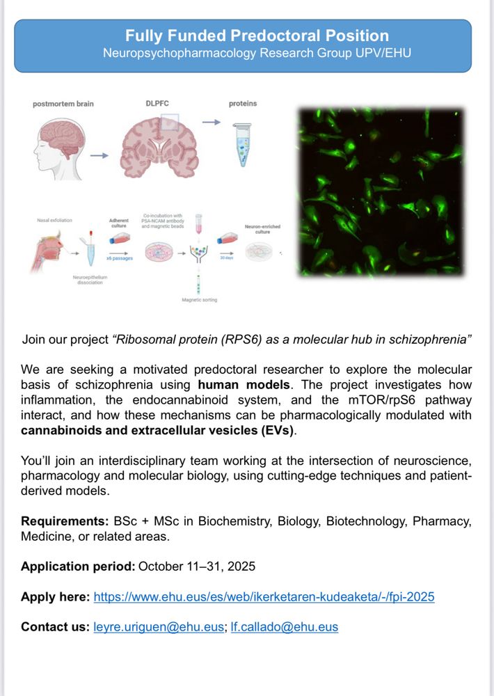
Job offer! Join our lab in the beautiful Basque Country! 🌊🌍🧠📚👇🏻
October 23, 2025 at 12:19 PM
Job offer! Join our lab in the beautiful Basque Country! 🌊🌍🧠📚👇🏻
Reposted by Vanja Tepavcevic

If you think your field needs a collection of curated reviews and protocols, let me know! Let's create it and help the community.
I'm always looking for ideas for CSH Protocols.
If you know of a field that would benefit from a curated and authoritative collection of review articles and step-by-step protocols, please let me know! 🧬🧪
We've recently done one on fly neurobiology, bacterial genetics, xenopus, and more!
If you know of a field that would benefit from a curated and authoritative collection of review articles and step-by-step protocols, please let me know! 🧬🧪
We've recently done one on fly neurobiology, bacterial genetics, xenopus, and more!

October 21, 2025 at 1:05 PM
If you think your field needs a collection of curated reviews and protocols, let me know! Let's create it and help the community.
Reposted by Vanja Tepavcevic
Mark your calendars!
The 28th Meeting of the French Glial Cell Club will take place Nov 17–20, 2026, in Sète.
🔹 Plenary lectures by Fiona Doetsch & Leda Dimou
🔹 5 symposia on glial diversity, astrocytes, human glia, EVs & modeling
👉 Full program available on our website: frenchglialcellclub.fr
The 28th Meeting of the French Glial Cell Club will take place Nov 17–20, 2026, in Sète.
🔹 Plenary lectures by Fiona Doetsch & Leda Dimou
🔹 5 symposia on glial diversity, astrocytes, human glia, EVs & modeling
👉 Full program available on our website: frenchglialcellclub.fr
October 20, 2025 at 2:38 PM
Mark your calendars!
The 28th Meeting of the French Glial Cell Club will take place Nov 17–20, 2026, in Sète.
🔹 Plenary lectures by Fiona Doetsch & Leda Dimou
🔹 5 symposia on glial diversity, astrocytes, human glia, EVs & modeling
👉 Full program available on our website: frenchglialcellclub.fr
The 28th Meeting of the French Glial Cell Club will take place Nov 17–20, 2026, in Sète.
🔹 Plenary lectures by Fiona Doetsch & Leda Dimou
🔹 5 symposia on glial diversity, astrocytes, human glia, EVs & modeling
👉 Full program available on our website: frenchglialcellclub.fr
Reposted by Vanja Tepavcevic
WHITE MATTER IN FOCUS SYMPOSIUM: EXPLORING THE ROLE OF MYELIN AND OLIGODENDROCYTES IN NEURODEGENERATION
neuroscience.cam.ac.uk/event-posts/...
neuroscience.cam.ac.uk/event-posts/...

White matter in focus symposium: Exploring the role of myelin and oligodendrocytes in neurodegeneration
Current research is increasingly highlighting the role of oligodendrocytes in neurodegeneration and specifically in dementia. Explore this theme across disease areas in the upcoming symposium White…
neuroscience.cam.ac.uk
October 20, 2025 at 4:31 PM
WHITE MATTER IN FOCUS SYMPOSIUM: EXPLORING THE ROLE OF MYELIN AND OLIGODENDROCYTES IN NEURODEGENERATION
neuroscience.cam.ac.uk/event-posts/...
neuroscience.cam.ac.uk/event-posts/...
Reposted by Vanja Tepavcevic

The microscopy core at Miami University is searching for a new director. Please consider sharing this job posting!
miamioh.wd5.myworkdayjobs.com/en-US/miamio...
miamioh.wd5.myworkdayjobs.com/en-US/miamio...
Director of the Center for Advanced Microscopy and Imaging
Job Title Director of the Center for Advanced Microscopy and Imaging Department Biology Department JM Worker Type Regular Pay Type Salary Position Salary Minimum $60,000 Position Salary Maximum $75,00...
miamioh.wd5.myworkdayjobs.com
October 17, 2025 at 12:45 AM
The microscopy core at Miami University is searching for a new director. Please consider sharing this job posting!
miamioh.wd5.myworkdayjobs.com/en-US/miamio...
miamioh.wd5.myworkdayjobs.com/en-US/miamio...
Reposted by Vanja Tepavcevic
Men’s brains shrink faster than women’s: what that means for Alzheimer’s nature.com/articles/d41...
#Alzheimers #dementia #health #wellness #science
#Alzheimers #dementia #health #wellness #science

Men’s brains shrink faster than women’s: what that means for Alzheimer’s
Women’s brains age more slowly, but that doesn’t seem to protect them from a common form of dementia.
nature.com
October 17, 2025 at 11:57 AM
Men’s brains shrink faster than women’s: what that means for Alzheimer’s nature.com/articles/d41...
#Alzheimers #dementia #health #wellness #science
#Alzheimers #dementia #health #wellness #science
Reposted by Vanja Tepavcevic
🧠 The Lipid #Brain Atlas is out now! If you think #lipids are boring and membranes are all the same, prepare to be surprised. Led by @lucafusarbassini.bsky.social with Giovanni D'Angelo's lab, we mapped membrane lipids in the mouse brain at high resolution.
www.biorxiv.org/cgi/content/...
www.biorxiv.org/cgi/content/...

October 16, 2025 at 6:23 AM
🧠 The Lipid #Brain Atlas is out now! If you think #lipids are boring and membranes are all the same, prepare to be surprised. Led by @lucafusarbassini.bsky.social with Giovanni D'Angelo's lab, we mapped membrane lipids in the mouse brain at high resolution.
www.biorxiv.org/cgi/content/...
www.biorxiv.org/cgi/content/...
Reposted by Vanja Tepavcevic
Interested in the heterogeneity of blood vessels in the CNS? Here we review the unique features of the spinal cord vasculature, its barrier properties and neurovascular interactions, highlighting unknown aspects and future research directions.
@CRC1366
@erc.europa.eu @immunosens.bsky.social
@CRC1366
@erc.europa.eu @immunosens.bsky.social

October 14, 2025 at 5:16 PM
Interested in the heterogeneity of blood vessels in the CNS? Here we review the unique features of the spinal cord vasculature, its barrier properties and neurovascular interactions, highlighting unknown aspects and future research directions.
@CRC1366
@erc.europa.eu @immunosens.bsky.social
@CRC1366
@erc.europa.eu @immunosens.bsky.social
Reposted by Vanja Tepavcevic
“Using Human Brain Tissue in Research”
NC3Rs supported @nc3rs.bsky.social
Date & time: Friday, 7 November 2025 13:00–16:00
Prof Maria Thom
Mr Martin Gillies
Mr Ciaran S. Hill
Location: UCL Institute of Advanced Studies
Registration (event is free): www.eventbrite.co.uk/e/networking...
NC3Rs supported @nc3rs.bsky.social
Date & time: Friday, 7 November 2025 13:00–16:00
Prof Maria Thom
Mr Martin Gillies
Mr Ciaran S. Hill
Location: UCL Institute of Advanced Studies
Registration (event is free): www.eventbrite.co.uk/e/networking...
Networking Event: Using Human Brain Tissue in Research
Networking event connecting researchers, clinicians & neurosurgeons to discuss practical and ethical aspects of human brain tissue research
www.eventbrite.co.uk
October 9, 2025 at 8:47 AM
“Using Human Brain Tissue in Research”
NC3Rs supported @nc3rs.bsky.social
Date & time: Friday, 7 November 2025 13:00–16:00
Prof Maria Thom
Mr Martin Gillies
Mr Ciaran S. Hill
Location: UCL Institute of Advanced Studies
Registration (event is free): www.eventbrite.co.uk/e/networking...
NC3Rs supported @nc3rs.bsky.social
Date & time: Friday, 7 November 2025 13:00–16:00
Prof Maria Thom
Mr Martin Gillies
Mr Ciaran S. Hill
Location: UCL Institute of Advanced Studies
Registration (event is free): www.eventbrite.co.uk/e/networking...
Another evidence for lactate influx, not efflux, in neurons. However, it suggests that in neonates, where levels of circulating lactate are high, lactate used by neurons is blood-derived, and that neonatal astrocytes might also use lactate.
www.nature.com/articles/s41...
www.nature.com/articles/s41...

Neuronal MCT2 promotes angiogenesis via lactate in the developing mouse neocortex - Cell Death & Differentiation
Cell Death & Differentiation - Neuronal MCT2 promotes angiogenesis via lactate in the developing mouse neocortex
www.nature.com
October 10, 2025 at 12:09 PM
Another evidence for lactate influx, not efflux, in neurons. However, it suggests that in neonates, where levels of circulating lactate are high, lactate used by neurons is blood-derived, and that neonatal astrocytes might also use lactate.
www.nature.com/articles/s41...
www.nature.com/articles/s41...
Reposted by Vanja Tepavcevic

Prenatal Acetaminophen Exposure Does Not Disrupt Human Fetal Brain Development in Cortical Organoid Models https://www.biorxiv.org/content/10.1101/2025.10.07.681041v1
October 8, 2025 at 7:15 PM
Prenatal Acetaminophen Exposure Does Not Disrupt Human Fetal Brain Development in Cortical Organoid Models https://www.biorxiv.org/content/10.1101/2025.10.07.681041v1
Alkalinization of glial cells increases lifespan and healthspan in C. Elegans
www.science.org/doi/10.1126/...
www.science.org/doi/10.1126/...

The loss of a ClC anion channel increases life span, health span, and stress resistance by alkalinizing a pair of glial cells in C. elegans
Alkalinization of glial cells mediates longevity, stress resistance, and protection from polyQ aggregation in C. elegans.
www.science.org
October 8, 2025 at 7:11 PM
Alkalinization of glial cells increases lifespan and healthspan in C. Elegans
www.science.org/doi/10.1126/...
www.science.org/doi/10.1126/...
Reposted by Vanja Tepavcevic

Immune signatures link myelin oligodendrocyte glycoprotein antibody–associated disease to other autoantibody-mediated conditions
www.science.org/doi/10.1126/...
www.science.org/doi/10.1126/...

Immune signatures link myelin oligodendrocyte glycoprotein antibody–associated disease to other autoantibody-mediated conditions
Single-cell immunoprofiling uncovered systemic immune cell signatures of neurological MOG antibody–associated disease.
www.science.org
October 8, 2025 at 7:04 PM
Immune signatures link myelin oligodendrocyte glycoprotein antibody–associated disease to other autoantibody-mediated conditions
www.science.org/doi/10.1126/...
www.science.org/doi/10.1126/...
Reposted by Vanja Tepavcevic
🚨Join our next @DEMONNetworkUK #Genetics and #Omics meeting on THURSDAY 9th October at 2.30 PM (UK Time). Delighted to have Kate Fodder as invited speaker – “Early #oligodendrocyte dysfunction signature in #Alzheimer’s disease: Insights from DNA #methylomics and #transcriptomics”.
DM for more info
DM for more info
October 8, 2025 at 2:57 PM
Reposted by Vanja Tepavcevic
Myelin pathology is a key feature of X-linked Dystonia Parkinsonism https://www.biorxiv.org/content/10.1101/2025.10.07.680990v1
October 8, 2025 at 1:15 PM
Myelin pathology is a key feature of X-linked Dystonia Parkinsonism https://www.biorxiv.org/content/10.1101/2025.10.07.680990v1
Reposted by Vanja Tepavcevic
GlialCAM Cytoplasmic Signaling in Oligodendrocytes and Astrocytes is Essential for White Matter Homeostasis in the Brain https://www.biorxiv.org/content/10.1101/2025.10.03.680331v1
October 4, 2025 at 8:15 AM
GlialCAM Cytoplasmic Signaling in Oligodendrocytes and Astrocytes is Essential for White Matter Homeostasis in the Brain https://www.biorxiv.org/content/10.1101/2025.10.03.680331v1
Reposted by Vanja Tepavcevic
SV2A-PET imaging uncovers cortical synapse loss in multiple sclerosis | Science Translational Medicine www.science.org/doi/10.1126/...

SV2A-PET imaging uncovers cortical synapse loss in multiple sclerosis
SV2A-PET reveals cortical synapse loss in multiple sclerosis (MS), highlighting a potential approach for unmasking gray matter pathology.
www.science.org
October 3, 2025 at 1:52 PM
SV2A-PET imaging uncovers cortical synapse loss in multiple sclerosis | Science Translational Medicine www.science.org/doi/10.1126/...