@builab.bsky.social
150 followers 120 following 130 posts
Posts Media Videos Starter Packs
builab.bsky.social
We also analysed SPEF1-KO cilia by cryo-ET to show that SPEF1 restricts C1 and C2 microtubule movement.
builab.bsky.social
In the final version, we included a lot more data and analysis compared to the previous biorxiv version. We achieved a 4.6 Angstrom resolution of the microtubule by subtomogram averaging of MT-SPEF1 complexes.
builab.bsky.social
Our newest paper is now online in Current Biology. In short, we identified 7 proteins in the tip of the cilia and found SPEF1 is a seam-binding protein and important for cilia stability. Great work from my postdoc Thibault Legal and everyone involved!

www.sciencedirect.com/science/arti...
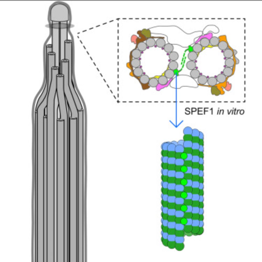
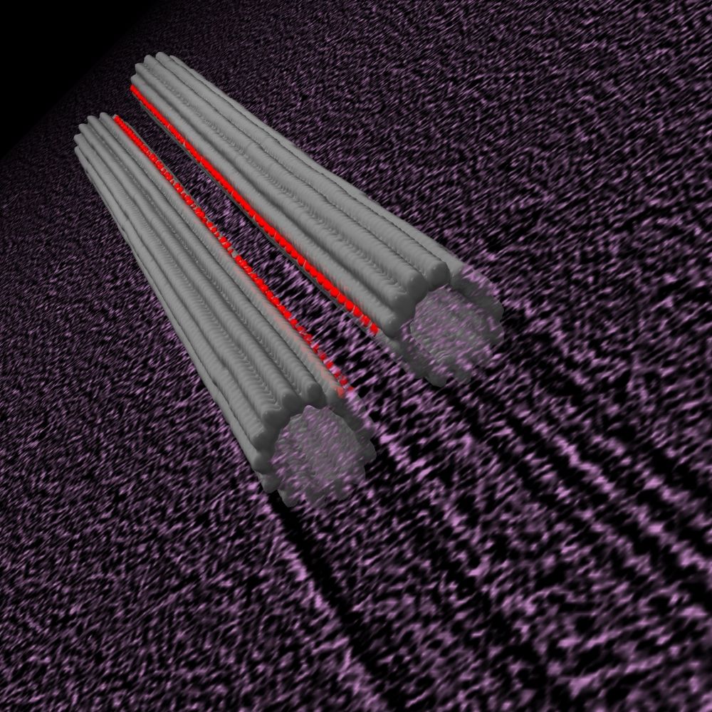
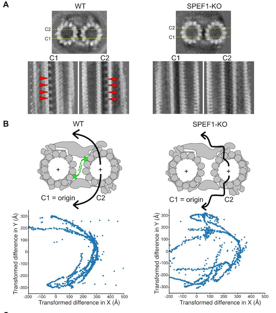
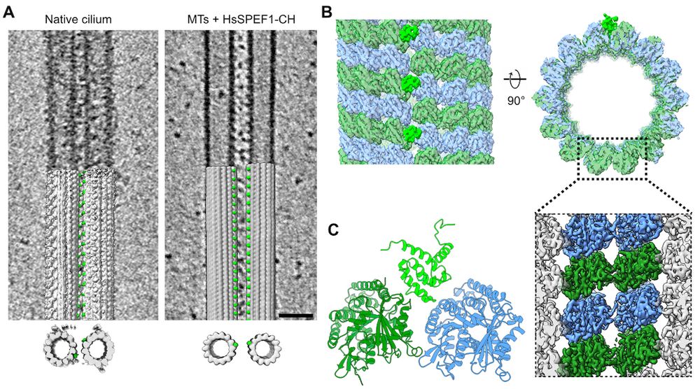
Structure of the ciliary tip central pair reveals the unique role of the microtubule-seam binding protein SPEF1
Motile cilia are unique organelles with the ability to move autonomously. The force generated by beating cilia propels cells and moves fluids. The cil…
www.sciencedirect.com
builab.bsky.social
This seems great. I want to working on our picking using AI in the summer. Perhaps, I can ask you for an installation and usage guide if I have problems.
builab.bsky.social
I hated Amira with the $$. Need to try DragonFly but most of our processing is done on the cluster so anything using command line is a big advantage. Perhaps, my summer project :)
builab.bsky.social
interesting I want to use dragonfly for segmentation but can we run dragonfly in command line for batch mode? Do you have a script to cluster and turn dragon fly into filament light?
builab.bsky.social
Very nice preprint from the Wallingford lab. Seems like there is a common theme of tip proteins as these proteins seem to be in the tip of the ciliate cilia too.
builab.bsky.social
Thanks. I found template matching is too slow for my need as I can manual pick microtubules much faster and more accurate. These tryout takes time so I 'd love to see people's experiences.
builab.bsky.social
#TeamTomo What is the open-source and good cryoET deep-picking program?
Reposted
embojournal.org
Cryo-electron tomography of Tetrahymena #cilia by Sam Li, David Agard, Mark Winey provides insights into the intricate spatial organization of basal body inner junctions, compromised by loss of Poc1
www.embopress.org/doi/full/10....
Reposted
pengxinchai.bsky.social
Excited to share our work on bioRxiv before my thesis defense! Together with my undergraduate mentee Diego, postdocs Wan and Jun, and my advisor Kai, we identified a novel dynein heavy chain subfamily, DNAHX, from sea urchin sperm axoneme.

Check out the details here: www.biorxiv.org/content/10.1...
DNAHX: a novel, non-motile dynein heavy chain subfamily, identified by cryo-EM endogenously
Ciliogenesis and cilia motility rely on the coordinated actions of diverse dyneins, yet the complexity of these motor proteins in cilia has posed challenges for understanding their specific roles. Tra...
www.biorxiv.org
Reposted
ruizhangmt.bsky.social
Best way to start 2025! Thrilled to share our Nature paper about the structures of axonemal components from sperm flagella and from epithelial cilia of the oviduct and brain ventricles. Great collaboration with Tzviya Zeev-Ben-Mordehai and @alanbrownhms.bsky.social

www.nature.com/articles/s41...
builab.bsky.social
Amazing work. It is so useful for us working with Tetrahymena and for cilia in general.
Reposted
computingcaitie.bsky.social
I am excited to share some of my PhD work on the motile cilia interactome revealed by XL/MS with @jbwallingford.bsky.social @edwardmarcotte.bsky.social Ophelia Papoulas, Chanjae Lee, David Taylor, and @builab.bsky.social www.cell.com/developmenta...
Reposted
chimerax.ucsf.edu
ChimeraX 1.9 has been released and is available now on our website
Download UCSF ChimeraX
www.cgl.ucsf.edu
builab.bsky.social
Thanks. Warp on Linux is awesome. This is my Twitter import so it is very old tweet
builab.bsky.social
My last post on Twitter before.
builab.bsky.social
Proudly present our new preprint on my first post here. We performed SPA on the ciliary tip and identify 7 proteins. Among them, we found the conserved protein SPEF1 at the microtubule seam. In vitro assays show that SPEF1 binds to the seam and stablizes microtubules.
www.biorxiv.org/content/10.1...
builab.bsky.social
The Bui lab Twitter will stop and we are migrating to Mastodon. Anyway, the last post is to thank the @Beck_Laboratory and @jankosinski @aobarska for the new wall art decoration. NPC rules :
builab.bsky.social
Our paper on the T- and N-pilus structure with the @ChristianBaron and @NatalieZeytuni labs is finally online. Great work from Amro Jaafar and @TheRealCSBlack and others.

authors.elsevier.com/a/1gElW3SNvc2%…
builab.bsky.social
Taking forever but finally here. We identified close to 40 MIPs in Tetrahymena doublet. Specifically, the MIPs at the outer junction (CFAP77, OJ2 & OJ3). @dynein_awk @TheRealCSBlack @ahmad__khalifa
builab.bsky.social
Just update the subtomo2Chimera withto visualize subtomo from Dynamo. Also, small fixes for relionsubtomo2ChimeraX. now you can also using stl file for visualization for more efficient memory #TeamTomo

github.com/builab/subtomo… dynamotable2ChimeraX.py
builab.bsky.social
#TeamTomo Which free software is good for segmentation of cryo-tomograms? Solutions for just 1 tomogram & many tomograms are welcome.
builab.bsky.social
Presenting the cryo-EM structures of T4SS T-pilus and N-pilus in a fruitful collaboration with @ChristianBaron @NatalieZeytuni @TheRealCSBlack and the twitterless Jaafar Amro. My first ever structures/publication on a bacterial protein

biorxiv.org/content/10.110…
Cryo-EM structure of the Agrobacterium tumefaciens T-pilus reveals the importance of positive charges in the lumen
Agrobacterium tumefaciens is a natural genetic engineer that transfers DNA into plants and this is the most frequently applied process for the generation of genetically modified plants. DNA transfer is mediated by a type IV secretion system localized in the cell envelope and extracellular T-pili. We here report the cryo-electron microscopic structures of the T-pilus at 3.2Å resolution and that of the related plasmid pKM101-determined N-pilus at 3Å resolution. Both pili contain a main pilus protein (VirB2 in A. tumefaciens and TraM in pKM101) and phospholipids arranged in a 5-start helical assembly. They contain positively charged amino acids in the pilus lumen and the lipids are positively charged in the T-pilus (phosphatidylcholine) conferring overall positive charge to the lumen. Mutagenesis of the lumen-exposed Arg91 residue in VirB2 resulted in protein destabilization and loss of pilus formation. Our results reveal that different phospholipids can be incorporated into type IV secretion system pili and that the charge of the lumen is of functional importance. ### Competing Interest Statement The authors have declared no competing interest.
www.biorxiv.org