


Hao Yin
@haoyin.bsky.social
Twitter/X @haoyin20
Vascular biologist
Vascular biologist
Pinned

“Time is brain” for cell therapies?🧠
Our #AdvSci perspective😆
@rrust.bsky.social
@dominikusbrian.com
@lydenlab.bsky.social
#StemCellTherapy for #Stroke should be timed to a biomarker-defined “window of receptivity”, not just the thrombolysis clock
advanced.onlinelibrary.wiley.com/doi/10.1002/...
Our #AdvSci perspective😆
@rrust.bsky.social
@dominikusbrian.com
@lydenlab.bsky.social
#StemCellTherapy for #Stroke should be timed to a biomarker-defined “window of receptivity”, not just the thrombolysis clock
advanced.onlinelibrary.wiley.com/doi/10.1002/...
Reposted by Hao Yin
This historic picture of Jupiter’s southern half always stops me in my tracks.
Voyager 2 grabbed it on 25 June 1979 when it was already down to 12 million kilometres and still closing fast.
photojournal.jpl.nasa.gov/catalog/PIA0...
Pic by NASA/JPL
🔭 🧪 #science
1/4
Voyager 2 grabbed it on 25 June 1979 when it was already down to 12 million kilometres and still closing fast.
photojournal.jpl.nasa.gov/catalog/PIA0...
Pic by NASA/JPL
🔭 🧪 #science
1/4

November 25, 2025 at 11:43 PM
This historic picture of Jupiter’s southern half always stops me in my tracks.
Voyager 2 grabbed it on 25 June 1979 when it was already down to 12 million kilometres and still closing fast.
photojournal.jpl.nasa.gov/catalog/PIA0...
Pic by NASA/JPL
🔭 🧪 #science
1/4
Voyager 2 grabbed it on 25 June 1979 when it was already down to 12 million kilometres and still closing fast.
photojournal.jpl.nasa.gov/catalog/PIA0...
Pic by NASA/JPL
🔭 🧪 #science
1/4
Reposted by Hao Yin
From Jupiter it went on to Saturn (1981), Uranus (1986), Neptune (1989).
On 5 Nov. 2018 it slipped out of the Sun’s domain into true interstellar space.
Right now, Nov. 2025, it’s some 141.49 AU away — 21.17 billion km — the farthest thing humans have ever sent, and it’s still talking to us.
4/4
On 5 Nov. 2018 it slipped out of the Sun’s domain into true interstellar space.
Right now, Nov. 2025, it’s some 141.49 AU away — 21.17 billion km — the farthest thing humans have ever sent, and it’s still talking to us.
4/4
November 25, 2025 at 11:43 PM
From Jupiter it went on to Saturn (1981), Uranus (1986), Neptune (1989).
On 5 Nov. 2018 it slipped out of the Sun’s domain into true interstellar space.
Right now, Nov. 2025, it’s some 141.49 AU away — 21.17 billion km — the farthest thing humans have ever sent, and it’s still talking to us.
4/4
On 5 Nov. 2018 it slipped out of the Sun’s domain into true interstellar space.
Right now, Nov. 2025, it’s some 141.49 AU away — 21.17 billion km — the farthest thing humans have ever sent, and it’s still talking to us.
4/4
Reposted by Hao Yin
So happy to announce our new preprint, “A geothermal amoeba sets a new upper temperature limit for eukaryotes.” We cultured a novel amoeba from Lassen Volcanic NP (CA, USA) that divides at 63°C (145°F) 🔥 - a new record for euk growth!
#protistsonsky 🧵
#protistsonsky 🧵
November 25, 2025 at 8:41 PM
So happy to announce our new preprint, “A geothermal amoeba sets a new upper temperature limit for eukaryotes.” We cultured a novel amoeba from Lassen Volcanic NP (CA, USA) that divides at 63°C (145°F) 🔥 - a new record for euk growth!
#protistsonsky 🧵
#protistsonsky 🧵
Downregulated FGF9 in type 2 diabetic #SkeletalMuscle
db/db🐭
Re-analysis of Human SkM #T2D #Sarcopenia transcriptomes
Hub genes: FGF9 BDH1 LDHA
#SciRep 2025
www.nature.com/articles/s41...
db/db🐭
Re-analysis of Human SkM #T2D #Sarcopenia transcriptomes
Hub genes: FGF9 BDH1 LDHA
#SciRep 2025
www.nature.com/articles/s41...

November 25, 2025 at 8:16 PM
Downregulated FGF9 in type 2 diabetic #SkeletalMuscle
db/db🐭
Re-analysis of Human SkM #T2D #Sarcopenia transcriptomes
Hub genes: FGF9 BDH1 LDHA
#SciRep 2025
www.nature.com/articles/s41...
db/db🐭
Re-analysis of Human SkM #T2D #Sarcopenia transcriptomes
Hub genes: FGF9 BDH1 LDHA
#SciRep 2025
www.nature.com/articles/s41...
Reposted by Hao Yin

Extracellular #RNAs play an important role in plants' communication with other organisms in their environment.
Read here about the secretion of extracellular (ex)RNA in plant–bacterial interaction.
🧪 #Science #Biology #Genetics
1- www.cell.com/trends/genet...
2- www.nature.com/articles/s41...
Read here about the secretion of extracellular (ex)RNA in plant–bacterial interaction.
🧪 #Science #Biology #Genetics
1- www.cell.com/trends/genet...
2- www.nature.com/articles/s41...

November 25, 2025 at 5:08 PM
Extracellular #RNAs play an important role in plants' communication with other organisms in their environment.
Read here about the secretion of extracellular (ex)RNA in plant–bacterial interaction.
🧪 #Science #Biology #Genetics
1- www.cell.com/trends/genet...
2- www.nature.com/articles/s41...
Read here about the secretion of extracellular (ex)RNA in plant–bacterial interaction.
🧪 #Science #Biology #Genetics
1- www.cell.com/trends/genet...
2- www.nature.com/articles/s41...
Reposted by Hao Yin

Looking forward to chairing a session with @doreenlau.bsky.social at the British Society for Immunology Congress next week!
Great line-up of speakers. Send an email if you would like to meet at or around the conference.
Great line-up of speakers. Send an email if you would like to meet at or around the conference.

November 25, 2025 at 3:48 PM
Looking forward to chairing a session with @doreenlau.bsky.social at the British Society for Immunology Congress next week!
Great line-up of speakers. Send an email if you would like to meet at or around the conference.
Great line-up of speakers. Send an email if you would like to meet at or around the conference.
Reposted by Hao Yin
Super proud to be part of SFB1348 — and excited to share that I now officially have my own lab in Münster! 🎉🪰
We’ll study #cellbio & #morphogenesis, focusing on organ sculpting and #chirality in #Drosophila.
I’ll soon hire my first #PhD student — feel free to reach out!
#juniorPI #Science #devbio
We’ll study #cellbio & #morphogenesis, focusing on organ sculpting and #chirality in #Drosophila.
I’ll soon hire my first #PhD student — feel free to reach out!
#juniorPI #Science #devbio

November 25, 2025 at 3:57 PM
Super proud to be part of SFB1348 — and excited to share that I now officially have my own lab in Münster! 🎉🪰
We’ll study #cellbio & #morphogenesis, focusing on organ sculpting and #chirality in #Drosophila.
I’ll soon hire my first #PhD student — feel free to reach out!
#juniorPI #Science #devbio
We’ll study #cellbio & #morphogenesis, focusing on organ sculpting and #chirality in #Drosophila.
I’ll soon hire my first #PhD student — feel free to reach out!
#juniorPI #Science #devbio
Reposted by Hao Yin

Dysregulated calcium signaling contributes to Alzheimer’s, but the molecular details are unresolved.
Here, Lauren H. Sansing & team report on changes of GLO1 with age & its association with calcium leak in degeneration, using mouse and macaque models: insight.jci.org/articles/vie...
Here, Lauren H. Sansing & team report on changes of GLO1 with age & its association with calcium leak in degeneration, using mouse and macaque models: insight.jci.org/articles/vie...
November 25, 2025 at 2:02 PM
Dysregulated calcium signaling contributes to Alzheimer’s, but the molecular details are unresolved.
Here, Lauren H. Sansing & team report on changes of GLO1 with age & its association with calcium leak in degeneration, using mouse and macaque models: insight.jci.org/articles/vie...
Here, Lauren H. Sansing & team report on changes of GLO1 with age & its association with calcium leak in degeneration, using mouse and macaque models: insight.jci.org/articles/vie...
Reposted by Hao Yin

🫁 The lungs are rarely the full story.
In rare diseases, the lungs often reveal what’s happening across the entire body, from molecular disruption to systemic complications.
Join Prof Dr Allan Lawrie (Imperial College London) and Dr Sterghios Moschos (Pulmobiomed).
🔗 Listen: youtu.be/8CQN9U2h388
In rare diseases, the lungs often reveal what’s happening across the entire body, from molecular disruption to systemic complications.
Join Prof Dr Allan Lawrie (Imperial College London) and Dr Sterghios Moschos (Pulmobiomed).
🔗 Listen: youtu.be/8CQN9U2h388
November 25, 2025 at 11:31 AM
🫁 The lungs are rarely the full story.
In rare diseases, the lungs often reveal what’s happening across the entire body, from molecular disruption to systemic complications.
Join Prof Dr Allan Lawrie (Imperial College London) and Dr Sterghios Moschos (Pulmobiomed).
🔗 Listen: youtu.be/8CQN9U2h388
In rare diseases, the lungs often reveal what’s happening across the entire body, from molecular disruption to systemic complications.
Join Prof Dr Allan Lawrie (Imperial College London) and Dr Sterghios Moschos (Pulmobiomed).
🔗 Listen: youtu.be/8CQN9U2h388
Reposted by Hao Yin
#DBfeature 🐠
A new powerful live imaging platform tracks Erk signalling dynamics in developing zebrafish hepatocytes at single-cell resolution.
By Faraz Ahmed Butt, Alessandro De Simone, Stefano Di Talia, Kenneth Poss
tinyurl.com/ve5vzp6c
A new powerful live imaging platform tracks Erk signalling dynamics in developing zebrafish hepatocytes at single-cell resolution.
By Faraz Ahmed Butt, Alessandro De Simone, Stefano Di Talia, Kenneth Poss
tinyurl.com/ve5vzp6c

November 25, 2025 at 12:35 PM
#DBfeature 🐠
A new powerful live imaging platform tracks Erk signalling dynamics in developing zebrafish hepatocytes at single-cell resolution.
By Faraz Ahmed Butt, Alessandro De Simone, Stefano Di Talia, Kenneth Poss
tinyurl.com/ve5vzp6c
A new powerful live imaging platform tracks Erk signalling dynamics in developing zebrafish hepatocytes at single-cell resolution.
By Faraz Ahmed Butt, Alessandro De Simone, Stefano Di Talia, Kenneth Poss
tinyurl.com/ve5vzp6c
Hao's Preprint Reading List
November 25th 2025
Ion channels in Lymphatic vessels
Liver inflammation in Pulmonary arterial hypertension
Perfusable Endometriosis on a chip
Piezo1, Angiopoietin 2, Schlemm's Canal
Spiral artery remodeling in human pregnancy
docs.google.com/document/d/e...
November 25th 2025
Ion channels in Lymphatic vessels
Liver inflammation in Pulmonary arterial hypertension
Perfusable Endometriosis on a chip
Piezo1, Angiopoietin 2, Schlemm's Canal
Spiral artery remodeling in human pregnancy
docs.google.com/document/d/e...

November 25, 2025 at 1:03 PM
Hao's Preprint Reading List
November 25th 2025
Ion channels in Lymphatic vessels
Liver inflammation in Pulmonary arterial hypertension
Perfusable Endometriosis on a chip
Piezo1, Angiopoietin 2, Schlemm's Canal
Spiral artery remodeling in human pregnancy
docs.google.com/document/d/e...
November 25th 2025
Ion channels in Lymphatic vessels
Liver inflammation in Pulmonary arterial hypertension
Perfusable Endometriosis on a chip
Piezo1, Angiopoietin 2, Schlemm's Canal
Spiral artery remodeling in human pregnancy
docs.google.com/document/d/e...
Reposted by Hao Yin
Still posting cytoskeleton videos, it seems. Actin this time.
Sample: Lifeact-eGFP in HeLa cells.
Modality: Airyscan confocal
Timestamp is mm:ss and the scale bar is 5 µm.
Sample: Lifeact-eGFP in HeLa cells.
Modality: Airyscan confocal
Timestamp is mm:ss and the scale bar is 5 µm.
November 23, 2025 at 3:21 AM
Still posting cytoskeleton videos, it seems. Actin this time.
Sample: Lifeact-eGFP in HeLa cells.
Modality: Airyscan confocal
Timestamp is mm:ss and the scale bar is 5 µm.
Sample: Lifeact-eGFP in HeLa cells.
Modality: Airyscan confocal
Timestamp is mm:ss and the scale bar is 5 µm.
Reposted by Hao Yin

It’s time to talk about epithelial geometry and cancer. How can the architecture of an epithelium affect how tumors will grow and spread? In this thread, @jorgealmagro.bsky.social
November 23, 2025 at 8:01 AM
It’s time to talk about epithelial geometry and cancer. How can the architecture of an epithelium affect how tumors will grow and spread? In this thread, @jorgealmagro.bsky.social
Reposted by Hao Yin
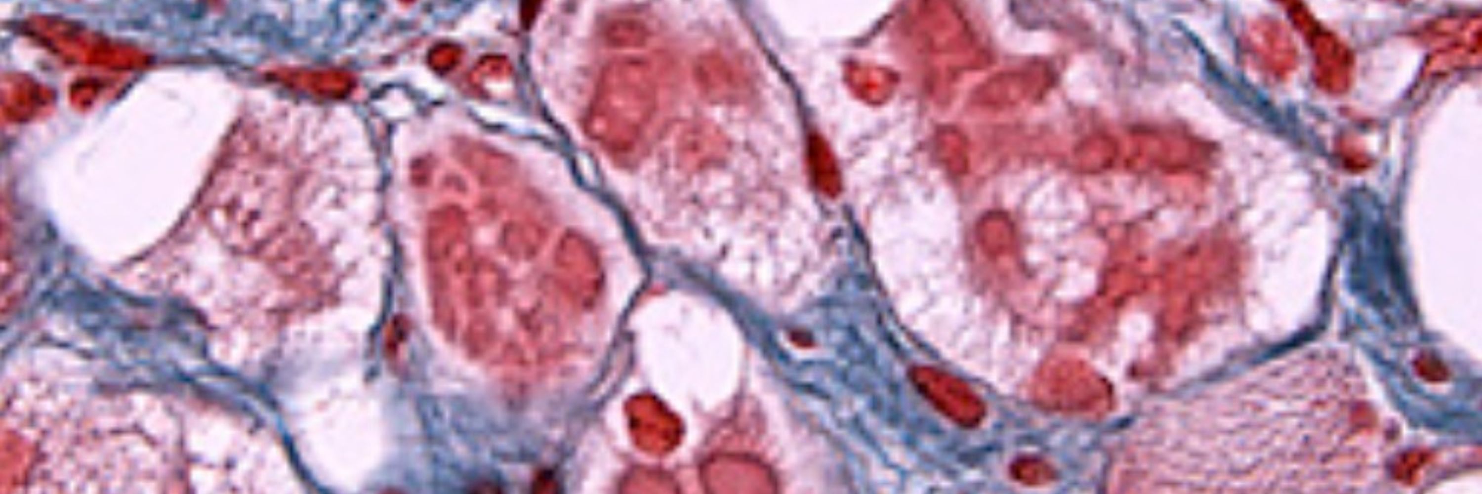
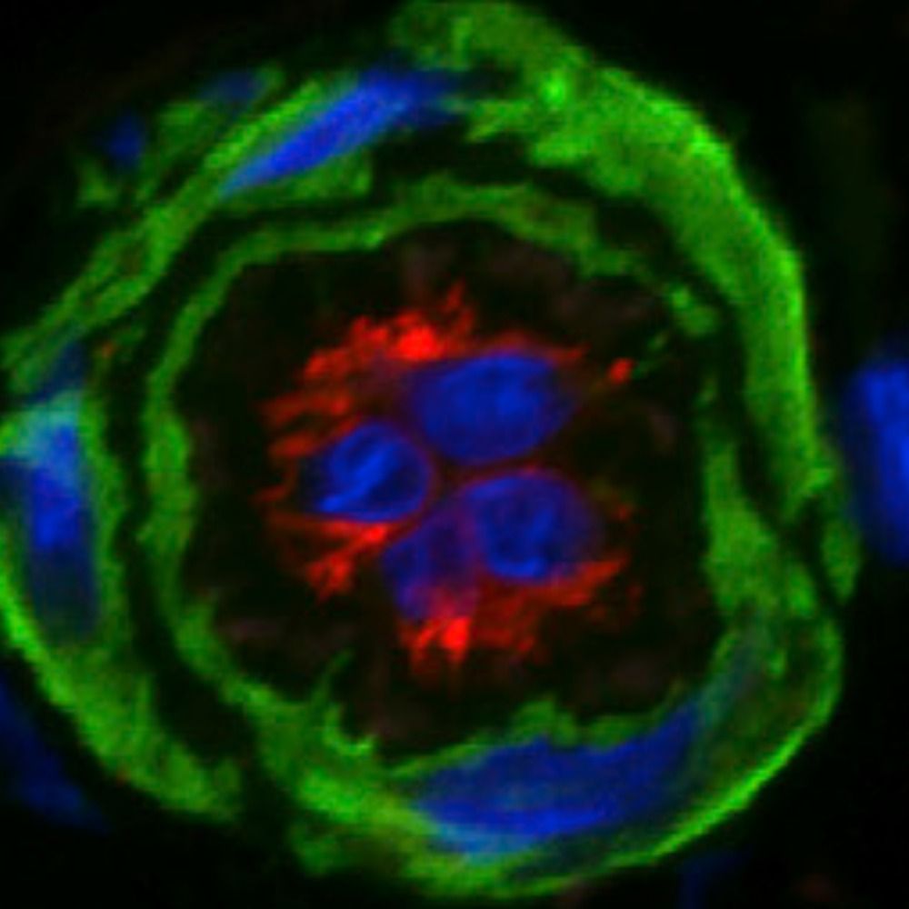
Gemini 3 created this figure in under 30 seconds from a couple reference images and a one sentence prompt
So I’m at a loss for words, honestly #MicroscopyMonday
So I’m at a loss for words, honestly #MicroscopyMonday

November 24, 2025 at 7:11 PM
Gemini 3 created this figure in under 30 seconds from a couple reference images and a one sentence prompt
So I’m at a loss for words, honestly #MicroscopyMonday
So I’m at a loss for words, honestly #MicroscopyMonday
Reposted by Hao Yin
Entire creation of new data that has no bearing upon any histological relevance. Remember LLM models are not open and we have no understanding of what NeuN, GFAP, or DAPI means in biological space within the LLM.
This is nonsense. But it looks at first blush as "real" to the uninformed.
Scary.
This is nonsense. But it looks at first blush as "real" to the uninformed.
Scary.
Gemini 3 created this figure in under 30 seconds from a couple reference images and a one sentence prompt
So I’m at a loss for words, honestly #MicroscopyMonday
So I’m at a loss for words, honestly #MicroscopyMonday
November 24, 2025 at 7:51 PM
Entire creation of new data that has no bearing upon any histological relevance. Remember LLM models are not open and we have no understanding of what NeuN, GFAP, or DAPI means in biological space within the LLM.
This is nonsense. But it looks at first blush as "real" to the uninformed.
Scary.
This is nonsense. But it looks at first blush as "real" to the uninformed.
Scary.
#MetaplasticBreastCancer #Immunotherapy
Pathological complete response was achieved in
1⃣2/33 Neoadjuvant Chemo
2⃣2/7 Neoadjuvant Chemo + Immunotherapy
Love to see some immunopathology on tumor-infiltrating lymphocytes & PD-L1
#BreastCancer 2025
www.dovepress.com/promising-re...
Pathological complete response was achieved in
1⃣2/33 Neoadjuvant Chemo
2⃣2/7 Neoadjuvant Chemo + Immunotherapy
Love to see some immunopathology on tumor-infiltrating lymphocytes & PD-L1
#BreastCancer 2025
www.dovepress.com/promising-re...

November 24, 2025 at 8:01 PM
#MetaplasticBreastCancer #Immunotherapy
Pathological complete response was achieved in
1⃣2/33 Neoadjuvant Chemo
2⃣2/7 Neoadjuvant Chemo + Immunotherapy
Love to see some immunopathology on tumor-infiltrating lymphocytes & PD-L1
#BreastCancer 2025
www.dovepress.com/promising-re...
Pathological complete response was achieved in
1⃣2/33 Neoadjuvant Chemo
2⃣2/7 Neoadjuvant Chemo + Immunotherapy
Love to see some immunopathology on tumor-infiltrating lymphocytes & PD-L1
#BreastCancer 2025
www.dovepress.com/promising-re...
Reposted by Hao Yin
This issue’s Cover: @ebrahim-lab.bsky.social @dominikrobak.bsky.social& team report on the septin cytoskeleton at apical junctions of intestinal epithelial cells, whereby it maintains barrier integrity & protects from inflammation: insight.jci.org/articles/vie...

November 24, 2025 at 5:06 PM
This issue’s Cover: @ebrahim-lab.bsky.social @dominikrobak.bsky.social& team report on the septin cytoskeleton at apical junctions of intestinal epithelial cells, whereby it maintains barrier integrity & protects from inflammation: insight.jci.org/articles/vie...
Reposted by Hao Yin
24 November 1859: 'On the Origin of Species', Charles Darwin's groundbreaking book, was published.
Darwin's magnum opus is considered to be the foundation of evolutionary biology and is undoubtedly one among the most important books in the history of science.
🧪 #science #histsci #Darwin
1/3
Darwin's magnum opus is considered to be the foundation of evolutionary biology and is undoubtedly one among the most important books in the history of science.
🧪 #science #histsci #Darwin
1/3

November 24, 2025 at 3:49 PM
Reposted by Hao Yin

This 🐍 venom made so many cool multi-day live imaging sessions possible 🔬🐟🩸 @chrmosimann.bsky.social @huiskenlab.bsky.social @daetwylerstephan.bsky.social

Synchronized development of zebrafish embryos immobilized by snake venom (alpha-bungarotoxin). Credit to Dr. Ian Swinburne. #ZebrafishZunday
November 23, 2025 at 9:29 PM
This 🐍 venom made so many cool multi-day live imaging sessions possible 🔬🐟🩸 @chrmosimann.bsky.social @huiskenlab.bsky.social @daetwylerstephan.bsky.social
u-Segment3D
Universal consensus 3D segmentation of cells from 2D segmented stacks
Work on complex networks, organelles & even endothelial filopodia🤯
A solid step toward single-cell 3D spatial omics 👹
@natmethods.nature.com 2025
www.nature.com/articles/s41...
Universal consensus 3D segmentation of cells from 2D segmented stacks
Work on complex networks, organelles & even endothelial filopodia🤯
A solid step toward single-cell 3D spatial omics 👹
@natmethods.nature.com 2025
www.nature.com/articles/s41...

November 24, 2025 at 12:44 PM
u-Segment3D
Universal consensus 3D segmentation of cells from 2D segmented stacks
Work on complex networks, organelles & even endothelial filopodia🤯
A solid step toward single-cell 3D spatial omics 👹
@natmethods.nature.com 2025
www.nature.com/articles/s41...
Universal consensus 3D segmentation of cells from 2D segmented stacks
Work on complex networks, organelles & even endothelial filopodia🤯
A solid step toward single-cell 3D spatial omics 👹
@natmethods.nature.com 2025
www.nature.com/articles/s41...
Single-nucleus profiling of Ossabaw pig🐷#Atherosclerosis model
Castrated♂️ 16-mo
+High fat & high fructose diet for 9 mo
Smooth muscle cell subclusters
⏬ACAA2+ SMC
⏬TRPC4+ SMC
⏫SPP1+ SMC
⏫NFATC2+ SMC
⏫Pericyte BDNF TGFβ Oncostatin M
⏫Myeloid SEMA3C
#iScience 2025
www.cell.com/iscience/ful...
Castrated♂️ 16-mo
+High fat & high fructose diet for 9 mo
Smooth muscle cell subclusters
⏬ACAA2+ SMC
⏬TRPC4+ SMC
⏫SPP1+ SMC
⏫NFATC2+ SMC
⏫Pericyte BDNF TGFβ Oncostatin M
⏫Myeloid SEMA3C
#iScience 2025
www.cell.com/iscience/ful...

November 23, 2025 at 8:00 PM
Single-nucleus profiling of Ossabaw pig🐷#Atherosclerosis model
Castrated♂️ 16-mo
+High fat & high fructose diet for 9 mo
Smooth muscle cell subclusters
⏬ACAA2+ SMC
⏬TRPC4+ SMC
⏫SPP1+ SMC
⏫NFATC2+ SMC
⏫Pericyte BDNF TGFβ Oncostatin M
⏫Myeloid SEMA3C
#iScience 2025
www.cell.com/iscience/ful...
Castrated♂️ 16-mo
+High fat & high fructose diet for 9 mo
Smooth muscle cell subclusters
⏬ACAA2+ SMC
⏬TRPC4+ SMC
⏫SPP1+ SMC
⏫NFATC2+ SMC
⏫Pericyte BDNF TGFβ Oncostatin M
⏫Myeloid SEMA3C
#iScience 2025
www.cell.com/iscience/ful...
Reposted by Hao Yin
miRNA have multiple functions beyond their role in modulating translation.

November 23, 2025 at 3:18 PM
miRNA have multiple functions beyond their role in modulating translation.
#RNAEditing #PulmonaryArterialHypertension
ADAR1 (both p150 & p110 isoforms) ⬇️in 👤PAH (in 🫁vascular smooth muscle cell)
SMC ADAR1 KO🐭 excerbates PAH
SMC ⏫dsRNA➡️⏫PKR-eIF2B-IFNβ➡️
Macrophage M1 polarization➡️
SMC proliferation
@kekeyuan.bsky.social #CircRes 2025
www.ahajournals.org/doi/10.1161/...
ADAR1 (both p150 & p110 isoforms) ⬇️in 👤PAH (in 🫁vascular smooth muscle cell)
SMC ADAR1 KO🐭 excerbates PAH
SMC ⏫dsRNA➡️⏫PKR-eIF2B-IFNβ➡️
Macrophage M1 polarization➡️
SMC proliferation
@kekeyuan.bsky.social #CircRes 2025
www.ahajournals.org/doi/10.1161/...

November 23, 2025 at 12:37 PM
#RNAEditing #PulmonaryArterialHypertension
ADAR1 (both p150 & p110 isoforms) ⬇️in 👤PAH (in 🫁vascular smooth muscle cell)
SMC ADAR1 KO🐭 excerbates PAH
SMC ⏫dsRNA➡️⏫PKR-eIF2B-IFNβ➡️
Macrophage M1 polarization➡️
SMC proliferation
@kekeyuan.bsky.social #CircRes 2025
www.ahajournals.org/doi/10.1161/...
ADAR1 (both p150 & p110 isoforms) ⬇️in 👤PAH (in 🫁vascular smooth muscle cell)
SMC ADAR1 KO🐭 excerbates PAH
SMC ⏫dsRNA➡️⏫PKR-eIF2B-IFNβ➡️
Macrophage M1 polarization➡️
SMC proliferation
@kekeyuan.bsky.social #CircRes 2025
www.ahajournals.org/doi/10.1161/...
Reposted by Hao Yin

🚀 Excited to launch a new series to keep #CardioObstetrics community updated with high-yield research in under 90 seconds.
“90-Second Cardio-OB Journal Scan”
with @cardioobdoc.bsky.social
Nov 14, 2025 article from @AJOG_thegray
🔗 www.ajog.org/article/S000...
#CardioSky #ACCRepOB #ACCCardioOB
“90-Second Cardio-OB Journal Scan”
with @cardioobdoc.bsky.social
Nov 14, 2025 article from @AJOG_thegray
🔗 www.ajog.org/article/S000...
#CardioSky #ACCRepOB #ACCCardioOB
November 14, 2025 at 2:23 PM
🚀 Excited to launch a new series to keep #CardioObstetrics community updated with high-yield research in under 90 seconds.
“90-Second Cardio-OB Journal Scan”
with @cardioobdoc.bsky.social
Nov 14, 2025 article from @AJOG_thegray
🔗 www.ajog.org/article/S000...
#CardioSky #ACCRepOB #ACCCardioOB
“90-Second Cardio-OB Journal Scan”
with @cardioobdoc.bsky.social
Nov 14, 2025 article from @AJOG_thegray
🔗 www.ajog.org/article/S000...
#CardioSky #ACCRepOB #ACCCardioOB
Reposted by Hao Yin
Attempt number seven at uploading this video of intermediate filaments in an enormous COS7 cell. I have a feeling the BlueSky compression will not do it any favors.
November 21, 2025 at 3:54 AM
Attempt number seven at uploading this video of intermediate filaments in an enormous COS7 cell. I have a feeling the BlueSky compression will not do it any favors.