


Miranda Marvel
@mmarvel.bsky.social
Researching the epigenetic regulation of vascular development in #zebrafish and #cavefish in the #WeinsteinLab
Pinned
Miranda Marvel
@mmarvel.bsky.social
· Apr 24

It's been a long time coming and I'm super excited to share our new preprint is out: "A novel transgenic reporter to study vertebrate epigenetics". We generated a zebrafish epigenetic reporter and found a fatty liver disease mutant with weird epigenetic phenotypes 👇
www.biorxiv.org/content/10.1...
www.biorxiv.org/content/10.1...
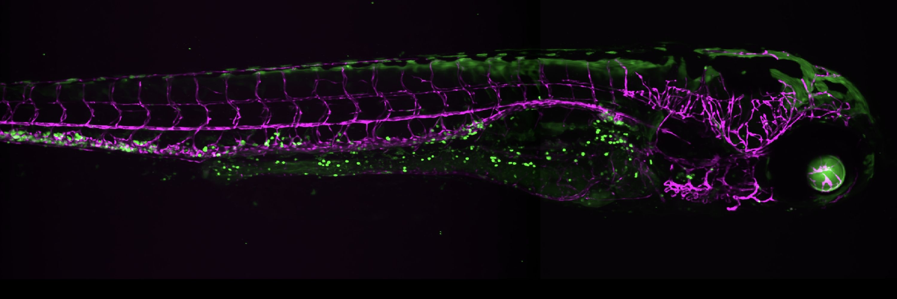
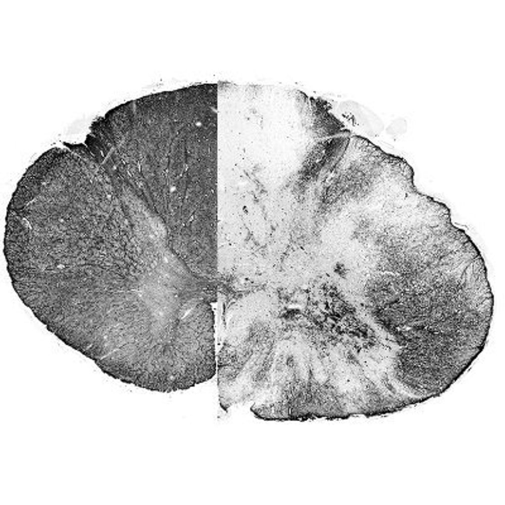
New preprint from the #WeinsteinLab is out with some awesome imaging of an important injury model! 🐟🧠 #zebrafish

Check out our recent preprint, "Live longitudinal imaging of meningeal cerebrovascular injury and its sequelae in adult zebrafish."
lnkd.in/gN7z6th4
lnkd.in/gN7z6th4
November 20, 2025 at 4:22 PM
New preprint from the #WeinsteinLab is out with some awesome imaging of an important injury model! 🐟🧠 #zebrafish
Reposted by Miranda Marvel

@jimmykjm.bsky.social and @isabellaclsci.bsky.social et al. focus on epigenetic changes during fin regeneration in this preprint:
www.biorxiv.org/content/10.1...
www.biorxiv.org/content/10.1...

Early epigenetic priming of regeneration studied with a novel transgenic epigenetic reporter
Tissue regeneration requires previously differentiated cells to regain developmental plasticity. However, the upstream mechanisms initiating this process remain poorly understood. Here, we leverage a ...
www.biorxiv.org
April 28, 2025 at 2:15 PM
@jimmykjm.bsky.social and @isabellaclsci.bsky.social et al. focus on epigenetic changes during fin regeneration in this preprint:
www.biorxiv.org/content/10.1...
www.biorxiv.org/content/10.1...
Reposted by Miranda Marvel
Two great new #Zebrafish preprints from the #WeinsteinLab using a new transgenic that reports epigenetic silencing and activation! Great to study epigenetic changes in vertebrates, and we even did an ENU screen. Excellent work by @mmarvel.bsky.social et al. 🧪
www.biorxiv.org/content/10.1...
www.biorxiv.org/content/10.1...
April 28, 2025 at 2:14 PM
Two great new #Zebrafish preprints from the #WeinsteinLab using a new transgenic that reports epigenetic silencing and activation! Great to study epigenetic changes in vertebrates, and we even did an ENU screen. Excellent work by @mmarvel.bsky.social et al. 🧪
www.biorxiv.org/content/10.1...
www.biorxiv.org/content/10.1...

In this very cool 1-2 punch from the Weinstein lab, start by reading Miranda & co’s awesome pre-print about a novel epigenetic reporter and follow it up with @jimmykjm.bsky.social’s pre-print using it to characterize epigenetic reprogramming during fin regeneration: www.biorxiv.org/content/10.1...
April 25, 2025 at 6:00 PM
It's been a long time coming and I'm super excited to share our new preprint is out: "A novel transgenic reporter to study vertebrate epigenetics". We generated a zebrafish epigenetic reporter and found a fatty liver disease mutant with weird epigenetic phenotypes 👇
www.biorxiv.org/content/10.1...
www.biorxiv.org/content/10.1...
April 24, 2025 at 10:42 PM
It's been a long time coming and I'm super excited to share our new preprint is out: "A novel transgenic reporter to study vertebrate epigenetics". We generated a zebrafish epigenetic reporter and found a fatty liver disease mutant with weird epigenetic phenotypes 👇
www.biorxiv.org/content/10.1...
www.biorxiv.org/content/10.1...
Reposted by Miranda Marvel
Happy to share a new preprint from my lab! We characterize the on/off kinetics, light dosage-dependence, and more for a suite of optogenetic signaling activators in zebrafish embryos 💡 www.biorxiv.org/content/10.1...

An optogenetic toolkit for robust activation of FGF, BMP, and Nodal signaling in zebrafish
Cell signaling regulates a wide range of biological processes including development, homeostasis, and disease. Accessible technologies to precisely manipulate signaling have important applications in ...
www.biorxiv.org
April 21, 2025 at 1:47 PM
Happy to share a new preprint from my lab! We characterize the on/off kinetics, light dosage-dependence, and more for a suite of optogenetic signaling activators in zebrafish embryos 💡 www.biorxiv.org/content/10.1...
Reposted by Miranda Marvel
Great new work led by @biomarina-vg.bsky.social on zebrafish meninges!
Particularly 🤩 is the descriptions of ependymin-expressing meningeal cells (not a fibroblast!) & ablation studies showing their essential role in viability. So much more to learn!
Congrats Marina & team on this great work! 🦓🐟🧠
Particularly 🤩 is the descriptions of ependymin-expressing meningeal cells (not a fibroblast!) & ablation studies showing their essential role in viability. So much more to learn!
Congrats Marina & team on this great work! 🦓🐟🧠


April 20, 2025 at 2:40 PM
Great new work led by @biomarina-vg.bsky.social on zebrafish meninges!
Particularly 🤩 is the descriptions of ependymin-expressing meningeal cells (not a fibroblast!) & ablation studies showing their essential role in viability. So much more to learn!
Congrats Marina & team on this great work! 🦓🐟🧠
Particularly 🤩 is the descriptions of ependymin-expressing meningeal cells (not a fibroblast!) & ablation studies showing their essential role in viability. So much more to learn!
Congrats Marina & team on this great work! 🦓🐟🧠
Reposted by Miranda Marvel
We recently described the Axillary Lymphoid Organ (ALO) in the #Zebrafish. It's on the OUTSIDE of the fish, making it great for imaging! Learn more by checking out our paper in @jem.org rupress.org/jem/article-...
April 3, 2025 at 12:07 AM
We recently described the Axillary Lymphoid Organ (ALO) in the #Zebrafish. It's on the OUTSIDE of the fish, making it great for imaging! Learn more by checking out our paper in @jem.org rupress.org/jem/article-...
Reposted by Miranda Marvel
Excited to see our work describing an external immune organ in the #zebrafish published in JEM! Big thanks to Aurora Kraus and @zebrafishmk.bsky.social and all of our other amazing collaborators including @edanfoley.bsky.social @biomarina-vg.bsky.social #WeinsteinLab rupress.org/jem/article-... 🧪
April 1, 2025 at 11:38 PM
Excited to see our work describing an external immune organ in the #zebrafish published in JEM! Big thanks to Aurora Kraus and @zebrafishmk.bsky.social and all of our other amazing collaborators including @edanfoley.bsky.social @biomarina-vg.bsky.social #WeinsteinLab rupress.org/jem/article-... 🧪
Reposted by Miranda Marvel

New: @zebrafish007.bsky.social, @zebrafishmk.bsky.social et al. #WeinsteinLab describe a previously uncharacterized external, experimentally accessible secondary lymphoid organ in the #zebrafish located above the pectoral fin. rupress.org/jem/article/...
April 1, 2025 at 4:55 PM
New: @zebrafish007.bsky.social, @zebrafishmk.bsky.social et al. #WeinsteinLab describe a previously uncharacterized external, experimentally accessible secondary lymphoid organ in the #zebrafish located above the pectoral fin. rupress.org/jem/article/...
Well deserved recognition 🎉
Special congrats to NIH Senior Investigator Brant Weinstein, Ph.D., who was named a fellow of the American Association for the Advancement of Science. Dr. Weinstein was honored for his contributions to vascular biology. Learn more at go.nih.gov/sl2B1ka. #zebrafish @izfs.bsky.social

April 4, 2025 at 2:33 AM
Well deserved recognition 🎉
Reposted by Miranda Marvel

Exciting news! We’re thrilled to announce that tapir Yuna gave birth to a rare and endangered Malayan tapir calf Sunday night. The newborn, covered in distinctive white spots and stripes resembling a fuzzy walking watermelon, is only the second tapir born in our 120-year history.
February 4, 2025 at 7:00 PM
Exciting news! We’re thrilled to announce that tapir Yuna gave birth to a rare and endangered Malayan tapir calf Sunday night. The newborn, covered in distinctive white spots and stripes resembling a fuzzy walking watermelon, is only the second tapir born in our 120-year history.
Reposted by Miranda Marvel
A couple of weeks left to apply to join Development's *Pathway to Independence* programme
Support & mentoring for post-docs on the academic job market
Deadline: January 31
journals.biologists.com/dev/pages/pi...
Support & mentoring for post-docs on the academic job market
Deadline: January 31
journals.biologists.com/dev/pages/pi...
Development PI Programme | Development | The Company of Biologists
Development PI Programme | Development | The Company of Biologists
Development's Pathway to Independence programme
Development is excited to announce our second call for our Pathway to I...
journals.biologists.com
January 17, 2025 at 9:21 AM
A couple of weeks left to apply to join Development's *Pathway to Independence* programme
Support & mentoring for post-docs on the academic job market
Deadline: January 31
journals.biologists.com/dev/pages/pi...
Support & mentoring for post-docs on the academic job market
Deadline: January 31
journals.biologists.com/dev/pages/pi...
An amazing #zebrafish research position opportunity with a great lab at NIH, check this out if you're on the job market!
Great opportunity! Research position in the Sheppard Lab at the NIH in Bethesda. Zebrafish + imaging + lymphatic vessels = AWESOME! 🧪🔬🐟
careers.criver.com/job/Bethesda...
careers.criver.com/job/Bethesda...

January 15, 2025 at 3:17 PM
An amazing #zebrafish research position opportunity with a great lab at NIH, check this out if you're on the job market!
Reposted by Miranda Marvel
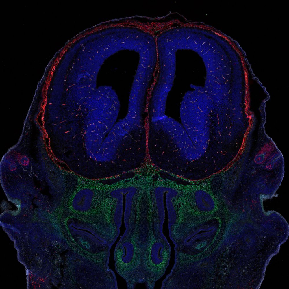
And somehow I missed it going live last week 😂 - our #zebrafish glial-vascular paper is available for those finterested! One of the best ways to end 2024 as a #newPI www.cell.com/iscience/ful...
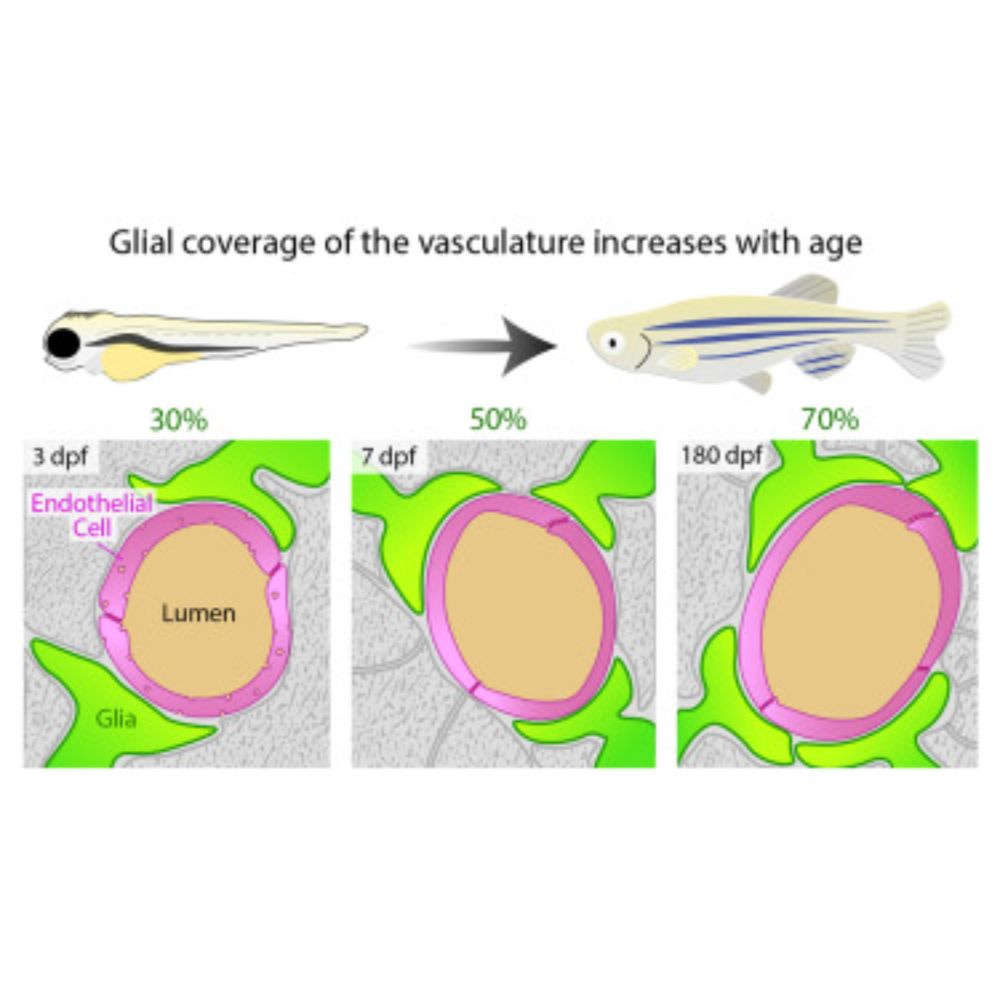
Zebrafish glial-vascular interactions progressively expand over the course of brain development
Glial-vascular interactions are critical for the formation and maintenance of brain blood vessels and the blood-brain barrier (BBB) in mammals, but their role in the zebrafish BBB remains unclear. Using three glial gene promoters—gfap, glast, and glastini (a truncated glast)—we explored glial-vascular development in zebrafish. Sparse labeling showed fewer glial-vascular interactions at early stages, with glial coverage and contact area increasing with age. Stable transgenic lines for glast and glastini revealed similar developmental increases, starting at ∼30% coverage at 3 days post-fertilization (dpf) and peaking at ∼60% by 10 dpf, and consistently higher glial coverage in the forebrain and midbrain than in the hindbrain.
www.cell.com
December 11, 2024 at 4:36 PM
And somehow I missed it going live last week 😂 - our #zebrafish glial-vascular paper is available for those finterested! One of the best ways to end 2024 as a #newPI www.cell.com/iscience/ful...
Reposted by Miranda Marvel

Out now in Dev Cell! scRNAseq trajectories describe the cascade of gene expression as cells differentiate, but turning them into a clear understanding of the underlying biology of a cell type's development remains challenging. authors.elsevier.com/c/1k9qj5Sx5g... (1/)

November 26, 2024 at 3:49 PM
Out now in Dev Cell! scRNAseq trajectories describe the cascade of gene expression as cells differentiate, but turning them into a clear understanding of the underlying biology of a cell type's development remains challenging. authors.elsevier.com/c/1k9qj5Sx5g... (1/)
Reposted by Miranda Marvel
#NewPI achievement unlocked: the first
#obrownlab publication is officially accepted (🍾) about #zebrafish #glia (green) and the vasculature (magenta). Thanks to our reviewers and congrats to all of the lab members! What a great 1st paper experience. Happy #fluorescencefriday ya'll!
#obrownlab publication is officially accepted (🍾) about #zebrafish #glia (green) and the vasculature (magenta). Thanks to our reviewers and congrats to all of the lab members! What a great 1st paper experience. Happy #fluorescencefriday ya'll!
November 22, 2024 at 8:12 PM
#NewPI achievement unlocked: the first
#obrownlab publication is officially accepted (🍾) about #zebrafish #glia (green) and the vasculature (magenta). Thanks to our reviewers and congrats to all of the lab members! What a great 1st paper experience. Happy #fluorescencefriday ya'll!
#obrownlab publication is officially accepted (🍾) about #zebrafish #glia (green) and the vasculature (magenta). Thanks to our reviewers and congrats to all of the lab members! What a great 1st paper experience. Happy #fluorescencefriday ya'll!
Check out this super talented researcher (and a great person to boot) who's on the job market! 👇

Just swam over to Bluesky and decided to dip my fin in the water. I am a Postdoc in the Weinstein lab and currently on the faculty job market. I developed an injury system to study cutaneous wound healing in adult zebrafish. Check out my movie showing neutrophil recruitment after injury.
November 20, 2024 at 10:47 PM
Check out this super talented researcher (and a great person to boot) who's on the job market! 👇
Reposted by Miranda Marvel

Latest preprint from the lab!
Vascular mural cells protect the adult brain from haemorrhage but do not control the blood-brain barrier in developing zebrafish
www.biorxiv.org/content/10.1...
Vascular mural cells protect the adult brain from haemorrhage but do not control the blood-brain barrier in developing zebrafish
www.biorxiv.org/content/10.1...
Vascular mural cells protect the adult brain from haemorrhage but do not control the blood-brain barrier in developing zebrafish
The blood-brain barrier (BBB) protects the brain from circulating metabolites and plays central roles in neurological diseases. Endothelial cells (ECs) of the BBB are enwrapped by mural cells includin...
www.biorxiv.org
November 20, 2024 at 6:23 AM
Latest preprint from the lab!
Vascular mural cells protect the adult brain from haemorrhage but do not control the blood-brain barrier in developing zebrafish
www.biorxiv.org/content/10.1...
Vascular mural cells protect the adult brain from haemorrhage but do not control the blood-brain barrier in developing zebrafish
www.biorxiv.org/content/10.1...
Reposted by Miranda Marvel
Bones, scales, and lymphatic vessels in juvenile #Zebrafish for #FluorescenceFriday #WeinsteinLab 🧪🔬

November 15, 2024 at 2:29 PM
Bones, scales, and lymphatic vessels in juvenile #Zebrafish for #FluorescenceFriday #WeinsteinLab 🧪🔬
Reposted by Miranda Marvel
🥳 First Yaniv Lab paper to appear on this new platform!! Thanks Baptiste for the shout-out! 🎉

November 16, 2024 at 9:47 AM
🥳 First Yaniv Lab paper to appear on this new platform!! Thanks Baptiste for the shout-out! 🎉
Reposted by Miranda Marvel
Axons ❤️ ECM

November 16, 2024 at 2:06 PM
Axons ❤️ ECM
Reposted by Miranda Marvel
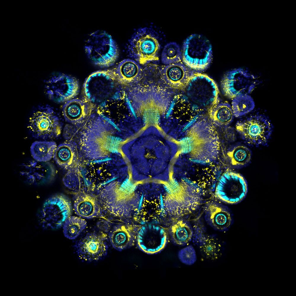
Shamelessly taking advantage of the bluesky migration to share again some of my favorite echinoderm pics 😀🤩
🔽 Acetylated tubulin (nerves and cilia) and phalloidin (muscles) stains in a juvenile sea urchin (Paracentrotus lividus) just after metamorphosis ⭐
🔽 Acetylated tubulin (nerves and cilia) and phalloidin (muscles) stains in a juvenile sea urchin (Paracentrotus lividus) just after metamorphosis ⭐
November 17, 2024 at 6:40 PM
Shamelessly taking advantage of the bluesky migration to share again some of my favorite echinoderm pics 😀🤩
🔽 Acetylated tubulin (nerves and cilia) and phalloidin (muscles) stains in a juvenile sea urchin (Paracentrotus lividus) just after metamorphosis ⭐
🔽 Acetylated tubulin (nerves and cilia) and phalloidin (muscles) stains in a juvenile sea urchin (Paracentrotus lividus) just after metamorphosis ⭐
Reposted by Miranda Marvel

And here’s the rest! A list of ECRs in the #zebrafish community 😎 Reach out if you think you should be added 🙏 🧪 go.bsky.app/HwxuHey
November 12, 2024 at 7:18 AM
And here’s the rest! A list of ECRs in the #zebrafish community 😎 Reach out if you think you should be added 🙏 🧪 go.bsky.app/HwxuHey
Reposted by Miranda Marvel
We joined the trend of Starter Packs! Check out our (non-exhaustive) list of Group Leaders & Senior Scientists that work with our favorite fishes! #zebrafish go.bsky.app/2cHYybY 🧪
November 11, 2024 at 9:45 AM
We joined the trend of Starter Packs! Check out our (non-exhaustive) list of Group Leaders & Senior Scientists that work with our favorite fishes! #zebrafish go.bsky.app/2cHYybY 🧪