


CellComM Lab
@cellcommlab.bsky.social
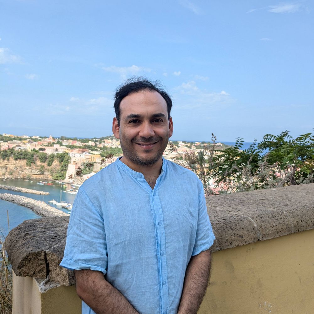
𝗖𝗲𝗹𝗹 𝗖𝗼𝗺𝘮𝘶𝘯𝘪𝘤𝘢𝘵𝘪𝘰𝘯 & 𝗠𝘪𝘨𝘳𝘢𝘵𝘪𝘰𝘯 Lab, headed by
@pjsaez.bsky.social #UKE_HH
We ❤ #CellComm #CellMigration 🧪🔬 tinyurl.com/5n8sss49
@pjsaez.bsky.social #UKE_HH
We ❤ #CellComm #CellMigration 🧪🔬 tinyurl.com/5n8sss49
Pinned
CellComM Lab
@cellcommlab.bsky.social
· Jan 24
#CellMigration for #FluorescenceFriday 🧪🔬
If a metastatic #cancer cell is offered 2 paths it decides very fast, bc it does matter where it goes...
OTOH, if a non-cancer #cell is offered 2 options it takes some time to explore and decide.
More in the paper: www.nature.com/articles/s41...
If a metastatic #cancer cell is offered 2 paths it decides very fast, bc it does matter where it goes...
OTOH, if a non-cancer #cell is offered 2 options it takes some time to explore and decide.
More in the paper: www.nature.com/articles/s41...
Reposted by CellComM Lab

JCS snapshot - Tracking coordinated cellular dynamics in time-lapse microscopy with ARCOS.px
@macdobry.bsky.social presents the key findings from their recent JCS paper with @bgraedel.bsky.social and colleagues @olivierpertz.bsky.social.
journals.biologists.com/jcs/article/...
@macdobry.bsky.social presents the key findings from their recent JCS paper with @bgraedel.bsky.social and colleagues @olivierpertz.bsky.social.
journals.biologists.com/jcs/article/...
January 29, 2026 at 2:24 PM
JCS snapshot - Tracking coordinated cellular dynamics in time-lapse microscopy with ARCOS.px
@macdobry.bsky.social presents the key findings from their recent JCS paper with @bgraedel.bsky.social and colleagues @olivierpertz.bsky.social.
journals.biologists.com/jcs/article/...
@macdobry.bsky.social presents the key findings from their recent JCS paper with @bgraedel.bsky.social and colleagues @olivierpertz.bsky.social.
journals.biologists.com/jcs/article/...
Reposted by CellComM Lab
Huge congrats to @manuelthery.bsky.social and all the Cytomorpho lab. The "Living Architectures" performance at @museeorsay.bsky.social was breathtakingly beautiful
January 25, 2026 at 9:22 AM
Huge congrats to @manuelthery.bsky.social and all the Cytomorpho lab. The "Living Architectures" performance at @museeorsay.bsky.social was breathtakingly beautiful
Reposted by CellComM Lab
Postdocs wanted - Please spread the word! We are looking for one bioinformatician and 1-2 cell biologists with different expertise! See the job ad below for more information and links to the job descriptions!

January 20, 2026 at 12:48 PM
Postdocs wanted - Please spread the word! We are looking for one bioinformatician and 1-2 cell biologists with different expertise! See the job ad below for more information and links to the job descriptions!
Reposted by CellComM Lab

279 postdoctoral fellowships!
Download freely our database of postdoctoral fellowships and grants. For each entry, we provide eligibility criteria, $ amount, deadline, etc.
Good luck!
Here: research.jhu.edu/rdt/funding-...
Download freely our database of postdoctoral fellowships and grants. For each entry, we provide eligibility criteria, $ amount, deadline, etc.
Good luck!
Here: research.jhu.edu/rdt/funding-...

January 20, 2026 at 1:22 PM
279 postdoctoral fellowships!
Download freely our database of postdoctoral fellowships and grants. For each entry, we provide eligibility criteria, $ amount, deadline, etc.
Good luck!
Here: research.jhu.edu/rdt/funding-...
Download freely our database of postdoctoral fellowships and grants. For each entry, we provide eligibility criteria, $ amount, deadline, etc.
Good luck!
Here: research.jhu.edu/rdt/funding-...
Reposted by CellComM Lab

Want to learn state-of-the-art deep learning techniques for microscopy image analysis? Apply by Jan. 15 to join us for a bootcamp at HHMI’s Janelia Research Campus. Gain hands-on experience with tools & frameworks you can use in your own research: bit.ly/4sqejiG 🧪

Deep Learning for Microscopy Image Analysis
Topics The following will be covered extensively during lectures, exercises, and project work: Image denoising and restoration (fully supervised and self-supervised) Image translation (e.g.,
bit.ly
January 13, 2026 at 9:33 PM
Want to learn state-of-the-art deep learning techniques for microscopy image analysis? Apply by Jan. 15 to join us for a bootcamp at HHMI’s Janelia Research Campus. Gain hands-on experience with tools & frameworks you can use in your own research: bit.ly/4sqejiG 🧪
Reposted by CellComM Lab

Our paper on the #DendroTweaks toolbox is now out as a Version of Record in @elife.bsky.social . doi.org/10.7554/eLif...
Find out more and try the toolbox online at dendrotweaks.dendrites.gr
Happy modeling!
#neuroskyence #compneurosky
Find out more and try the toolbox online at dendrotweaks.dendrites.gr
Happy modeling!
#neuroskyence #compneurosky

January 13, 2026 at 2:35 PM
Our paper on the #DendroTweaks toolbox is now out as a Version of Record in @elife.bsky.social . doi.org/10.7554/eLif...
Find out more and try the toolbox online at dendrotweaks.dendrites.gr
Happy modeling!
#neuroskyence #compneurosky
Find out more and try the toolbox online at dendrotweaks.dendrites.gr
Happy modeling!
#neuroskyence #compneurosky
Reposted by CellComM Lab
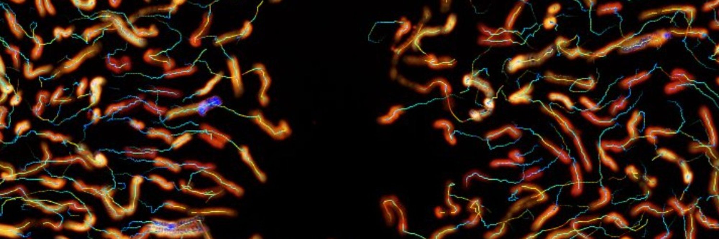
#FluorescenceFriday
Proud of our the last paper @cellcommlab.bsky.social & 1st one of @jboixcampos.bsky.social
We continue to study #CellMigration & how cells decide where to go: ↖️↗️⬇️?
(movie of the actin #cytoskeleton)
Full www.science.org/doi/10.1126/...
@focalplane.bsky.social @science.org 🧪🔬
Proud of our the last paper @cellcommlab.bsky.social & 1st one of @jboixcampos.bsky.social
We continue to study #CellMigration & how cells decide where to go: ↖️↗️⬇️?
(movie of the actin #cytoskeleton)
Full www.science.org/doi/10.1126/...
@focalplane.bsky.social @science.org 🧪🔬
January 9, 2026 at 4:23 PM
#FluorescenceFriday
Proud of our the last paper @cellcommlab.bsky.social & 1st one of @jboixcampos.bsky.social
We continue to study #CellMigration & how cells decide where to go: ↖️↗️⬇️?
(movie of the actin #cytoskeleton)
Full www.science.org/doi/10.1126/...
@focalplane.bsky.social @science.org 🧪🔬
Proud of our the last paper @cellcommlab.bsky.social & 1st one of @jboixcampos.bsky.social
We continue to study #CellMigration & how cells decide where to go: ↖️↗️⬇️?
(movie of the actin #cytoskeleton)
Full www.science.org/doi/10.1126/...
@focalplane.bsky.social @science.org 🧪🔬
Reposted by CellComM Lab
#HappyNewYear !
Let's start 2026 with a @cellcommlab.bsky.social paper
How many branches is OK during #CellMigration?
It depends on the #cell: for ex. immune cells are just fine.
In collab. w. #NirGov @hfspo.bsky.social
Full www.science.org/doi/10.1126/...
@focalplane.bsky.social @science.org 🧪🔬
Let's start 2026 with a @cellcommlab.bsky.social paper
How many branches is OK during #CellMigration?
It depends on the #cell: for ex. immune cells are just fine.
In collab. w. #NirGov @hfspo.bsky.social
Full www.science.org/doi/10.1126/...
@focalplane.bsky.social @science.org 🧪🔬
January 1, 2026 at 8:59 PM
#HappyNewYear !
Let's start 2026 with a @cellcommlab.bsky.social paper
How many branches is OK during #CellMigration?
It depends on the #cell: for ex. immune cells are just fine.
In collab. w. #NirGov @hfspo.bsky.social
Full www.science.org/doi/10.1126/...
@focalplane.bsky.social @science.org 🧪🔬
Let's start 2026 with a @cellcommlab.bsky.social paper
How many branches is OK during #CellMigration?
It depends on the #cell: for ex. immune cells are just fine.
In collab. w. #NirGov @hfspo.bsky.social
Full www.science.org/doi/10.1126/...
@focalplane.bsky.social @science.org 🧪🔬
Reposted by CellComM Lab

How do migrating cell groups balance force and flexibility?
Our new preprint shows that RhoGAP15B downregulates RhoA–Myosin activity to stabilise protrusions and enable efficient collective migration in vivo.
Happy New Year!
www.biorxiv.org/content/10.6...
Our new preprint shows that RhoGAP15B downregulates RhoA–Myosin activity to stabilise protrusions and enable efficient collective migration in vivo.
Happy New Year!
www.biorxiv.org/content/10.6...
December 31, 2025 at 2:14 PM
How do migrating cell groups balance force and flexibility?
Our new preprint shows that RhoGAP15B downregulates RhoA–Myosin activity to stabilise protrusions and enable efficient collective migration in vivo.
Happy New Year!
www.biorxiv.org/content/10.6...
Our new preprint shows that RhoGAP15B downregulates RhoA–Myosin activity to stabilise protrusions and enable efficient collective migration in vivo.
Happy New Year!
www.biorxiv.org/content/10.6...
Reposted by CellComM Lab

‼️🚨Hi all! The next Bioc Soc Membrane Contact Site Meeting will be in Chepstow, UK 28-30th Sept 2026. Fantastic speakers with talk slots still available. Sign up & abstract submission now open - limited spaces so register early! Hope to see you all there. Please share + RT 🙏

December 28, 2025 at 7:28 AM
‼️🚨Hi all! The next Bioc Soc Membrane Contact Site Meeting will be in Chepstow, UK 28-30th Sept 2026. Fantastic speakers with talk slots still available. Sign up & abstract submission now open - limited spaces so register early! Hope to see you all there. Please share + RT 🙏
Reposted by CellComM Lab
@biorxivpreprint.bsky.social CellTrap: A Microfluidic Platform Enabling Cell-Cell Interactions at Variable Effector to Target Ratios
www.biorxiv.org/content/10.6...
www.biorxiv.org/content/10.6...

December 27, 2025 at 5:34 PM
@biorxivpreprint.bsky.social CellTrap: A Microfluidic Platform Enabling Cell-Cell Interactions at Variable Effector to Target Ratios
www.biorxiv.org/content/10.6...
www.biorxiv.org/content/10.6...
Reposted by CellComM Lab
Malina Iwanski @mkiwanski.bsky.social, Lukas Kapitein and colleagues discover that stable microtubules reverse their polarity during neuronal development.
journals.biologists.com/jcs/article/...
#OpenAccess #ReadandPublish
journals.biologists.com/jcs/article/...
#OpenAccess #ReadandPublish
December 8, 2025 at 3:51 PM
Malina Iwanski @mkiwanski.bsky.social, Lukas Kapitein and colleagues discover that stable microtubules reverse their polarity during neuronal development.
journals.biologists.com/jcs/article/...
#OpenAccess #ReadandPublish
journals.biologists.com/jcs/article/...
#OpenAccess #ReadandPublish
Reposted by CellComM Lab

Do your samples move out of view? Tired of manually adjusting the stage? Introducing DySTrack developed by @zimengwu33.bsky.social and Jonas Hartmann from UCL, a tool that can be integrated into modern microscopes to automate the tracking of moving samples. #MicroscopyMonday doi.org/10.64898/202...
December 8, 2025 at 9:31 PM
Do your samples move out of view? Tired of manually adjusting the stage? Introducing DySTrack developed by @zimengwu33.bsky.social and Jonas Hartmann from UCL, a tool that can be integrated into modern microscopes to automate the tracking of moving samples. #MicroscopyMonday doi.org/10.64898/202...
Reposted by CellComM Lab
Interested in cell adhesion, evolution of multicellularity, or developing tools for emerging marine models?
My lab at UM is hiring a postdoc, and the application is now open:
🔗 tinyurl.com/28jvu4aa
If you know anyone looking for a postdoc, please pass this along!
My lab at UM is hiring a postdoc, and the application is now open:
🔗 tinyurl.com/28jvu4aa
If you know anyone looking for a postdoc, please pass this along!
December 8, 2025 at 6:37 PM
Interested in cell adhesion, evolution of multicellularity, or developing tools for emerging marine models?
My lab at UM is hiring a postdoc, and the application is now open:
🔗 tinyurl.com/28jvu4aa
If you know anyone looking for a postdoc, please pass this along!
My lab at UM is hiring a postdoc, and the application is now open:
🔗 tinyurl.com/28jvu4aa
If you know anyone looking for a postdoc, please pass this along!
Reposted by CellComM Lab
Come work with us! We are looking for a postdoc in #philbio or #philphysics to work on an interdisciplinary project that adopts the lens of self-organization & active matter to explore the boundary between living & nonliving systems www.kuleuven.be/personeel/jo... #academicsky #philjobs #HPS #evosky
December 8, 2025 at 11:21 AM
Come work with us! We are looking for a postdoc in #philbio or #philphysics to work on an interdisciplinary project that adopts the lens of self-organization & active matter to explore the boundary between living & nonliving systems www.kuleuven.be/personeel/jo... #academicsky #philjobs #HPS #evosky
Reposted by CellComM Lab

🔬 Call to create junior research groups at the Institut Pasteur
Focus: Infectious diseases, host-microbe interactions, vaccines
Special interest: AI methodologies
📅 Deadline: Feb 9, 2026
👥 2-12 years post-PhD
Apply now 📝 research.pasteur.fr/en/call/crea...
#JobOpportunity #Research
Focus: Infectious diseases, host-microbe interactions, vaccines
Special interest: AI methodologies
📅 Deadline: Feb 9, 2026
👥 2-12 years post-PhD
Apply now 📝 research.pasteur.fr/en/call/crea...
#JobOpportunity #Research

Creation of new junior research groups at the Institut Pasteur - Call for applications 2026 - Research
The Institut Pasteur is launching an international call to recruit new junior research group leaders leveraging cutting-edge transdisciplinary approaches to exploring infectious diseases, host-microbe...
research.pasteur.fr
December 8, 2025 at 8:53 AM
🔬 Call to create junior research groups at the Institut Pasteur
Focus: Infectious diseases, host-microbe interactions, vaccines
Special interest: AI methodologies
📅 Deadline: Feb 9, 2026
👥 2-12 years post-PhD
Apply now 📝 research.pasteur.fr/en/call/crea...
#JobOpportunity #Research
Focus: Infectious diseases, host-microbe interactions, vaccines
Special interest: AI methodologies
📅 Deadline: Feb 9, 2026
👥 2-12 years post-PhD
Apply now 📝 research.pasteur.fr/en/call/crea...
#JobOpportunity #Research
Reposted by CellComM Lab
Hot from the #preprint
Leukocyte + #CellMigration + physics = cool
Microtubules enable torque-based steering during dendritic cell migration.
Actomyosin contractility is required for noise-modulation strategy used by neutrophils.
Full @biorxivpreprint.bsky.social www.biorxiv.org/content/10.1...
Leukocyte + #CellMigration + physics = cool
Microtubules enable torque-based steering during dendritic cell migration.
Actomyosin contractility is required for noise-modulation strategy used by neutrophils.
Full @biorxivpreprint.bsky.social www.biorxiv.org/content/10.1...

Torque-based immune cell chemotaxis in complex environments
Directed migration in chemical gradients is crucial to the immune response, yet how immune cells navigate complex tissues remains incompletely understood. Using in vitro migration assays and theoretic...
www.biorxiv.org
November 23, 2025 at 2:39 PM
Hot from the #preprint
Leukocyte + #CellMigration + physics = cool
Microtubules enable torque-based steering during dendritic cell migration.
Actomyosin contractility is required for noise-modulation strategy used by neutrophils.
Full @biorxivpreprint.bsky.social www.biorxiv.org/content/10.1...
Leukocyte + #CellMigration + physics = cool
Microtubules enable torque-based steering during dendritic cell migration.
Actomyosin contractility is required for noise-modulation strategy used by neutrophils.
Full @biorxivpreprint.bsky.social www.biorxiv.org/content/10.1...
Reposted by CellComM Lab
Still posting cytoskeleton videos, it seems. Actin this time.
Sample: Lifeact-eGFP in HeLa cells.
Modality: Airyscan confocal
Timestamp is mm:ss and the scale bar is 5 µm.
Sample: Lifeact-eGFP in HeLa cells.
Modality: Airyscan confocal
Timestamp is mm:ss and the scale bar is 5 µm.
November 23, 2025 at 3:21 AM
Still posting cytoskeleton videos, it seems. Actin this time.
Sample: Lifeact-eGFP in HeLa cells.
Modality: Airyscan confocal
Timestamp is mm:ss and the scale bar is 5 µm.
Sample: Lifeact-eGFP in HeLa cells.
Modality: Airyscan confocal
Timestamp is mm:ss and the scale bar is 5 µm.
Reposted by CellComM Lab

It’s time to talk about epithelial geometry and cancer. How can the architecture of an epithelium affect how tumors will grow and spread? In this thread, @jorgealmagro.bsky.social
November 23, 2025 at 8:01 AM
It’s time to talk about epithelial geometry and cancer. How can the architecture of an epithelium affect how tumors will grow and spread? In this thread, @jorgealmagro.bsky.social
Reposted by CellComM Lab
Synchronized development of zebrafish embryos immobilized by snake venom (alpha-bungarotoxin). Credit to Dr. Ian Swinburne. #ZebrafishZunday
November 23, 2025 at 12:12 PM
Synchronized development of zebrafish embryos immobilized by snake venom (alpha-bungarotoxin). Credit to Dr. Ian Swinburne. #ZebrafishZunday
Reposted by CellComM Lab
Even if "provisionally" accepted, those are the best words/news I've heard for quite a while...
Here some #blebs to "provisionally" cellebrate.
Let's see this #cell dance under the microscope.
@focalplane.bsky.social #fluorescencefriday #microscopy 🧪🔬
Here some #blebs to "provisionally" cellebrate.
Let's see this #cell dance under the microscope.
@focalplane.bsky.social #fluorescencefriday #microscopy 🧪🔬
November 21, 2025 at 2:55 PM
Even if "provisionally" accepted, those are the best words/news I've heard for quite a while...
Here some #blebs to "provisionally" cellebrate.
Let's see this #cell dance under the microscope.
@focalplane.bsky.social #fluorescencefriday #microscopy 🧪🔬
Here some #blebs to "provisionally" cellebrate.
Let's see this #cell dance under the microscope.
@focalplane.bsky.social #fluorescencefriday #microscopy 🧪🔬
Reposted by CellComM Lab

I'm back at regulardly doing some live imaging so I have new little videos of actin in the immune synapse.
November 21, 2025 at 2:17 PM
I'm back at regulardly doing some live imaging so I have new little videos of actin in the immune synapse.
Reposted by CellComM Lab
The law of the jungle.
Interactions of cells in a collective lead to global rotation.
In 80% of the case HUVEC cells turn clockwise.
How many cells does it take for this to happen?
Interactions of cells in a collective lead to global rotation.
In 80% of the case HUVEC cells turn clockwise.
How many cells does it take for this to happen?
November 21, 2025 at 1:07 PM
The law of the jungle.
Interactions of cells in a collective lead to global rotation.
In 80% of the case HUVEC cells turn clockwise.
How many cells does it take for this to happen?
Interactions of cells in a collective lead to global rotation.
In 80% of the case HUVEC cells turn clockwise.
How many cells does it take for this to happen?
Reposted by CellComM Lab

#AsgardArchaea team, led by @archaeal.bsky.social fr @texasscience.bsky.social — sequencing the DNA collected fr mouth of Rio de la Plata to the continental shelf of Uruguay to detect Asgards, a group of single-celled organisms & our closest microbial relatives on the tree of life. bit.ly/3MdniTH
November 21, 2025 at 12:07 AM
#AsgardArchaea team, led by @archaeal.bsky.social fr @texasscience.bsky.social — sequencing the DNA collected fr mouth of Rio de la Plata to the continental shelf of Uruguay to detect Asgards, a group of single-celled organisms & our closest microbial relatives on the tree of life. bit.ly/3MdniTH