


Milena Schuhmacher
@mschuhmacher.bsky.social
ELISIR group leader at EPFL working on lipids and membranes using chemical biology
Reposted by Milena Schuhmacher
Registration for THE chemical biology conference of 2026 is now open! EMBO ChemBio 2026 in Heidelberg
DeGrado, Arikin, Picotti (Keynotes). @lmkdassama.bsky.social @brianliau.bsky.social @rhodamine110.bsky.social @benlehner.bsky.social @alitavassoli.bsky.social
www.embl.org/about/info/c...
DeGrado, Arikin, Picotti (Keynotes). @lmkdassama.bsky.social @brianliau.bsky.social @rhodamine110.bsky.social @benlehner.bsky.social @alitavassoli.bsky.social
www.embl.org/about/info/c...
Chemical biology 2026
www.embl.org
November 11, 2025 at 5:46 PM
Registration for THE chemical biology conference of 2026 is now open! EMBO ChemBio 2026 in Heidelberg
DeGrado, Arikin, Picotti (Keynotes). @lmkdassama.bsky.social @brianliau.bsky.social @rhodamine110.bsky.social @benlehner.bsky.social @alitavassoli.bsky.social
www.embl.org/about/info/c...
DeGrado, Arikin, Picotti (Keynotes). @lmkdassama.bsky.social @brianliau.bsky.social @rhodamine110.bsky.social @benlehner.bsky.social @alitavassoli.bsky.social
www.embl.org/about/info/c...
Fantastic story! Congratulations to everyone involved!
🧠 The Lipid #Brain Atlas is out now! If you think #lipids are boring and membranes are all the same, prepare to be surprised. Led by @lucafusarbassini.bsky.social with Giovanni D'Angelo's lab, we mapped membrane lipids in the mouse brain at high resolution.
www.biorxiv.org/cgi/content/...
www.biorxiv.org/cgi/content/...

October 16, 2025 at 3:49 PM
Fantastic story! Congratulations to everyone involved!
Reposted by Milena Schuhmacher

Registration open: 77. Mosbacher Kolloquium "More than lipidic barriers - New horizons in membrane biology" from March 25-28, 2026
lnkd.in/dgsW4c3
Scientific Organizers: Britta Brügger, Robert Ernst, André Nadler, Christian Ungermann
Early registration & poster abstracts until January 31, 2026
lnkd.in/dgsW4c3
Scientific Organizers: Britta Brügger, Robert Ernst, André Nadler, Christian Ungermann
Early registration & poster abstracts until January 31, 2026

October 9, 2025 at 6:25 AM
Registration open: 77. Mosbacher Kolloquium "More than lipidic barriers - New horizons in membrane biology" from March 25-28, 2026
lnkd.in/dgsW4c3
Scientific Organizers: Britta Brügger, Robert Ernst, André Nadler, Christian Ungermann
Early registration & poster abstracts until January 31, 2026
lnkd.in/dgsW4c3
Scientific Organizers: Britta Brügger, Robert Ernst, André Nadler, Christian Ungermann
Early registration & poster abstracts until January 31, 2026
Reposted by Milena Schuhmacher
Previous preprint is now published in RSC Chemical Biology.
If you are interested in plasma membrane labeling, see the paper! 👇
pubs.rsc.org/en/content/a...
If you are interested in plasma membrane labeling, see the paper! 👇
pubs.rsc.org/en/content/a...
September 8, 2025 at 8:02 AM
Previous preprint is now published in RSC Chemical Biology.
If you are interested in plasma membrane labeling, see the paper! 👇
pubs.rsc.org/en/content/a...
If you are interested in plasma membrane labeling, see the paper! 👇
pubs.rsc.org/en/content/a...
So honored to be awarded the ERC Starting Grant! I am very grateful for this support and looking forward to more exciting #lipidtime 🥳
Also, we will have positions available next year. So stay tuned!
Also, we will have positions available next year. So stay tuned!

📣 The ERC Starting Grant call results are out!
Find out which early-career researchers will receive funding this year, what they will be investigating, where they will be based... plus lots of other #ERCStG facts & figures for 2025!
➡️ buff.ly/IsafuFh
#FrontierResearch 🇪🇺#EUfunded #HorizonEurope
Find out which early-career researchers will receive funding this year, what they will be investigating, where they will be based... plus lots of other #ERCStG facts & figures for 2025!
➡️ buff.ly/IsafuFh
#FrontierResearch 🇪🇺#EUfunded #HorizonEurope
September 4, 2025 at 8:18 PM
So honored to be awarded the ERC Starting Grant! I am very grateful for this support and looking forward to more exciting #lipidtime 🥳
Also, we will have positions available next year. So stay tuned!
Also, we will have positions available next year. So stay tuned!
Reposted by Milena Schuhmacher

In the new book Labwork to Leadership, chemist Jen Heemstra offers actionable leadership advice to principal investigators.
Read the #ScienceBooks Review: https://scim.ag/4mljVr1
Read the #ScienceBooks Review: https://scim.ag/4mljVr1

August 19, 2025 at 6:49 PM
In the new book Labwork to Leadership, chemist Jen Heemstra offers actionable leadership advice to principal investigators.
Read the #ScienceBooks Review: https://scim.ag/4mljVr1
Read the #ScienceBooks Review: https://scim.ag/4mljVr1
Reposted by Milena Schuhmacher
Have you published lipid-protein interactome data? Please let us know, we’d love to include it in the repository. The goal is to build a centralized hub for the scientific community.
Huge thanks to Gaelen Guzman, a graduate student/postdoc in the lab who built it from scratch.
Huge thanks to Gaelen Guzman, a graduate student/postdoc in the lab who built it from scratch.
August 12, 2025 at 2:13 PM
Have you published lipid-protein interactome data? Please let us know, we’d love to include it in the repository. The goal is to build a centralized hub for the scientific community.
Huge thanks to Gaelen Guzman, a graduate student/postdoc in the lab who built it from scratch.
Huge thanks to Gaelen Guzman, a graduate student/postdoc in the lab who built it from scratch.
Reposted by Milena Schuhmacher
Many thanks Helge!
August 21, 2025 at 6:51 AM
Many thanks Helge!
We are hiring!
If you are looking for a postdoc position and you are interested in signaling lipids and proteomics, come join us in beautiful Switzerland!
More information on www.epfl.ch/labs/gr-schu...
If you are looking for a postdoc position and you are interested in signaling lipids and proteomics, come join us in beautiful Switzerland!
More information on www.epfl.ch/labs/gr-schu...

Open positions
GR-SCHUHMACHER
www.epfl.ch
August 12, 2025 at 9:32 AM
We are hiring!
If you are looking for a postdoc position and you are interested in signaling lipids and proteomics, come join us in beautiful Switzerland!
More information on www.epfl.ch/labs/gr-schu...
If you are looking for a postdoc position and you are interested in signaling lipids and proteomics, come join us in beautiful Switzerland!
More information on www.epfl.ch/labs/gr-schu...
Reposted by Milena Schuhmacher

Novel Research in Biological Artificial Intelligence: Expressions of interest and nominations for a Max Planck Director to lead research in the field of Biological Artificial Intelligence @mpi-cbg.de and @csbdresden.bsky.social. More information: www.mpi-cbg.de/news-outreac...
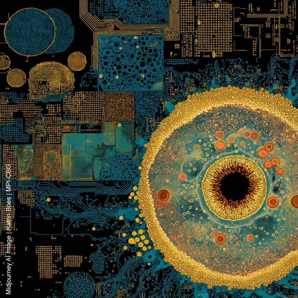
Novel Research in Biological Artificial Intelligence
Expressions of interest and nominations for a Max Planck Director to lead research in the field of Biological Artificial Intelligence.
www.mpi-cbg.de
June 26, 2025 at 11:26 AM
Novel Research in Biological Artificial Intelligence: Expressions of interest and nominations for a Max Planck Director to lead research in the field of Biological Artificial Intelligence @mpi-cbg.de and @csbdresden.bsky.social. More information: www.mpi-cbg.de/news-outreac...
Reposted by Milena Schuhmacher
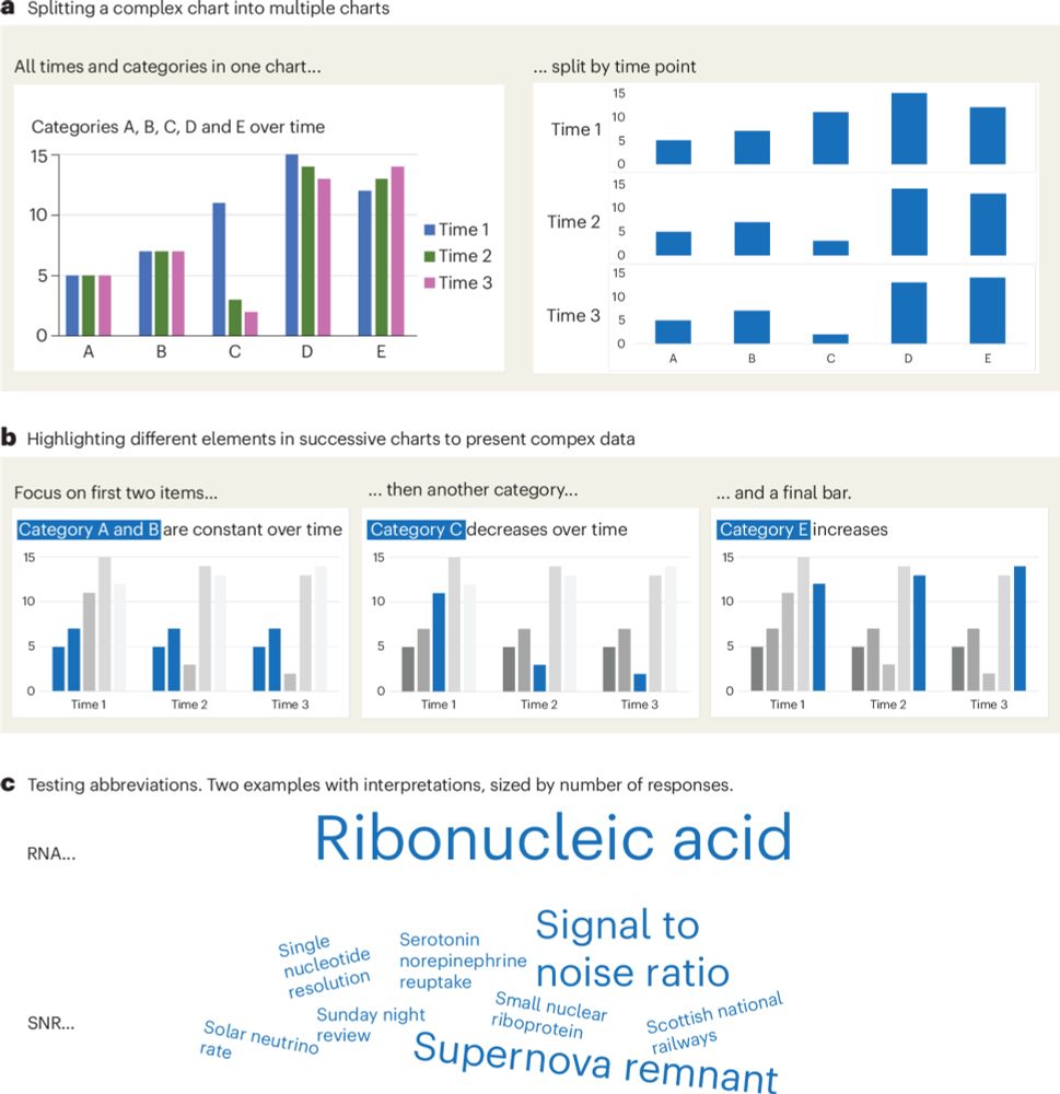
I gave in! After students asking for it, I now made a simple figure design checklist.
To help all scientists w/o graphic skills create clear, accessible, and truthful charts!
-> Out in @nature Cell Biology: rdcu.be/erwl4
#DataVisualization #PhD #SciComm
Thx for review @bethcimini.bsky.social + 2
To help all scientists w/o graphic skills create clear, accessible, and truthful charts!
-> Out in @nature Cell Biology: rdcu.be/erwl4
#DataVisualization #PhD #SciComm
Thx for review @bethcimini.bsky.social + 2
A checklist for designing and improving the visualization of scientific data
Nature Cell Biology - Creating clear and engaging scientific figures is crucial to communicate complex data. In this Comment, I condense principles from design, visual perception and data...
rdcu.be
June 18, 2025 at 8:33 AM
I gave in! After students asking for it, I now made a simple figure design checklist.
To help all scientists w/o graphic skills create clear, accessible, and truthful charts!
-> Out in @nature Cell Biology: rdcu.be/erwl4
#DataVisualization #PhD #SciComm
Thx for review @bethcimini.bsky.social + 2
To help all scientists w/o graphic skills create clear, accessible, and truthful charts!
-> Out in @nature Cell Biology: rdcu.be/erwl4
#DataVisualization #PhD #SciComm
Thx for review @bethcimini.bsky.social + 2
Reposted by Milena Schuhmacher

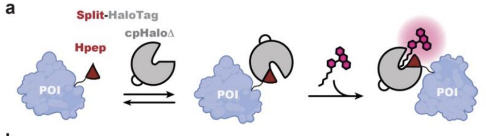
New preprint by @kjohnsson.bsky.social lab!
A new split Halotag system with higher affinity of the complements + lower background. The system works with our SiR-CA & CPY-CA halotag ligands and enables STED imaging or FLIM multiplexing.
The short 14 aa tag Hpep enables easy cloning free CRISPR-KI.
A new split Halotag system with higher affinity of the complements + lower background. The system works with our SiR-CA & CPY-CA halotag ligands and enables STED imaging or FLIM multiplexing.
The short 14 aa tag Hpep enables easy cloning free CRISPR-KI.

![Live-cell confocal imaging of histone H2B type-2E-Hpep11 CRISPR KI cell lines after 2-hours labeling with the specified CA- ligand [100 nM]. Images were taken with optimal image acquisition parameter for each dye. Scale bar: 50 μm.](https://cdn.bsky.app/img/feed_thumbnail/plain/did:plc:uf4m7zlmcjlg2biwn4s2k32y/bafkreibozn5fz6sgeksqrz3vmy6uiydtnvwmb5my7r3xecko2erokryn3a@jpeg)
![(a) Confocal laser scanning microscopy (CLSM) and STED images of mitochondria in U2OS cells coexpressing cpHaloΔ3 and TOM20-Hpep, either overexpressed or endogenously tagged. Scale bar: 1 μm. Pixel intensities scaled according to reference bar. (b) Representative CLSM, STED images of the CRISPR/Cas9 KI cells expressing TOM20 tagged with intact HaloTag (upper) or Hpep11 (bottom). (c) Intensity profiles along mitochondrial tubules (red and blue lines in b). Scale bar: 2 μm. (d) Representative CLSM and STED images of endogenous Hpep11-tagged clathrin with cpHaloΔ3 coexpression. Scale bar: 10 μm (overview) and 2 μm (magnification). (e) Representative CLSM, STED
images of endogenously tagged tubulin beta 4B with Hpep11. Scale bar: 10 μm (overview) and 2 μm (magnification). (f) Intensity profiles along tubulin filaments (red and blue lines in e) Means ± s.d. of the filament diameters were calculated as full width at half maximum (FWHM) from n=20 microtubule filaments, ≥ 2 images. A slight increase in cytosolic signal was noted in cells tagged with split-HaloTagat TUBB4B, compared to cells tagged with the full-length HaloTag, which may result from the presence of unbound but labeled cpHaloΔ3. All images were acquired after labeling with CA-SiR [100 nM] for one hour.](https://cdn.bsky.app/img/feed_thumbnail/plain/did:plc:uf4m7zlmcjlg2biwn4s2k32y/bafkreielty6fex37xy4caechbw5ebo3eypyiyvxxhkigeazd5gmy6tvlay@jpeg)
June 17, 2025 at 11:09 AM
New preprint by @kjohnsson.bsky.social lab!
A new split Halotag system with higher affinity of the complements + lower background. The system works with our SiR-CA & CPY-CA halotag ligands and enables STED imaging or FLIM multiplexing.
The short 14 aa tag Hpep enables easy cloning free CRISPR-KI.
A new split Halotag system with higher affinity of the complements + lower background. The system works with our SiR-CA & CPY-CA halotag ligands and enables STED imaging or FLIM multiplexing.
The short 14 aa tag Hpep enables easy cloning free CRISPR-KI.
Reposted by Milena Schuhmacher
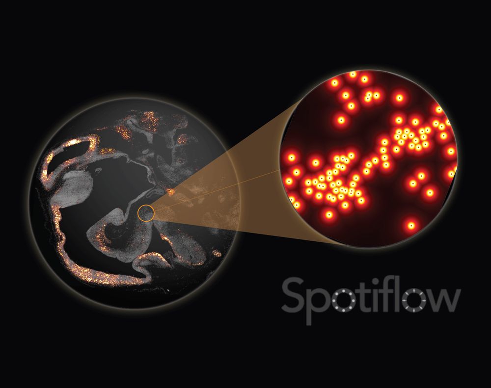
Out today in @natmethods.nature.com : Spotiflow, our transcript localization method for imaging-based spatial transcriptomics. Led by amazing PhD student @albertdm.bsky.social, joint work w @gioelelamanno.bsky.social at EPFL / @scadsai.bsky.social
www.nature.com/articles/s41...
rdcu.be/epIB7
www.nature.com/articles/s41...
rdcu.be/epIB7
June 6, 2025 at 7:06 PM
Out today in @natmethods.nature.com : Spotiflow, our transcript localization method for imaging-based spatial transcriptomics. Led by amazing PhD student @albertdm.bsky.social, joint work w @gioelelamanno.bsky.social at EPFL / @scadsai.bsky.social
www.nature.com/articles/s41...
rdcu.be/epIB7
www.nature.com/articles/s41...
rdcu.be/epIB7
Reposted by Milena Schuhmacher
90% of you probably don’t need to read this.
But maybe some of you are curious & 10% will feel seen. Or a little less alone. This isn’t about seeking sympathy.
It’s about sharing something hard to say out loud;
partly to heal, partly in case someone needs to hear it too. (1/3)
tinyurl.com/DudinO
But maybe some of you are curious & 10% will feel seen. Or a little less alone. This isn’t about seeking sympathy.
It’s about sharing something hard to say out loud;
partly to heal, partly in case someone needs to hear it too. (1/3)
tinyurl.com/DudinO
Omaya Dudin
Interview with Omaya Dudin, who uses Ichthyosporea as models to study how and why
unicellular organisms evolved multicellularity at the University of Geneva.
www.cell.com
June 10, 2025 at 9:41 AM
90% of you probably don’t need to read this.
But maybe some of you are curious & 10% will feel seen. Or a little less alone. This isn’t about seeking sympathy.
It’s about sharing something hard to say out loud;
partly to heal, partly in case someone needs to hear it too. (1/3)
tinyurl.com/DudinO
But maybe some of you are curious & 10% will feel seen. Or a little less alone. This isn’t about seeking sympathy.
It’s about sharing something hard to say out loud;
partly to heal, partly in case someone needs to hear it too. (1/3)
tinyurl.com/DudinO
Reposted by Milena Schuhmacher

Over 150 participants from 23 countries came to Varna, Bulgaria for the 2025 FEBS Special Meeting/13th ICC. Five days of outstanding science, networking, socializing, and community! We are grateful to all the sponsors and look forward to the next one!
June 1, 2025 at 7:38 PM
Over 150 participants from 23 countries came to Varna, Bulgaria for the 2025 FEBS Special Meeting/13th ICC. Five days of outstanding science, networking, socializing, and community! We are grateful to all the sponsors and look forward to the next one!
Reposted by Milena Schuhmacher
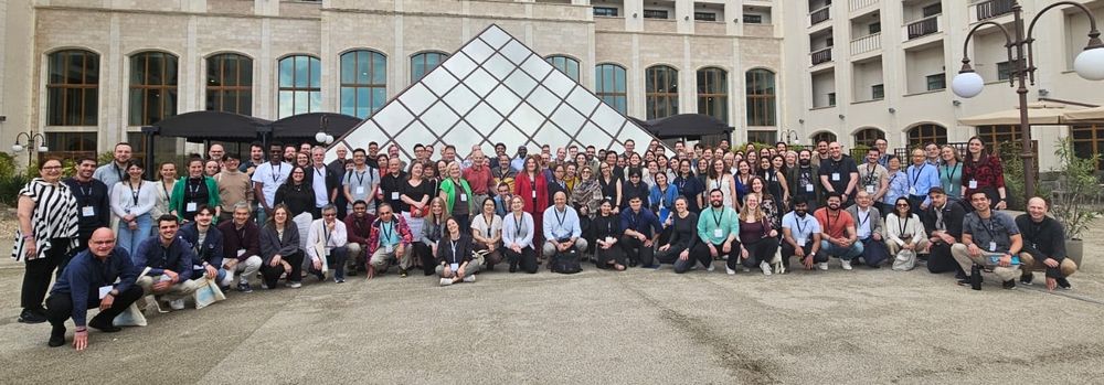
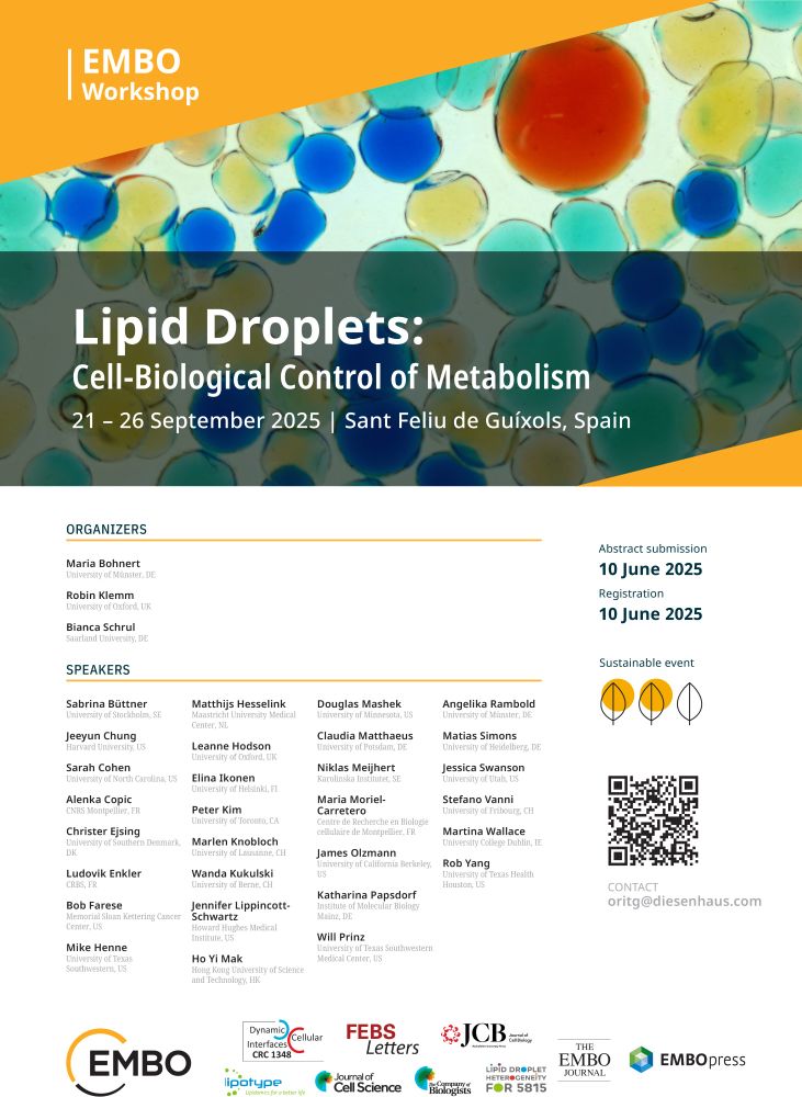
The abstract submission deadline for the 2nd edition of our #EMBOLipidDroplets Workshop (21-26 September 2025) is approaching.
Check out the agenda with an amazing lineup of speakers and submit your abstract before 10 June 2025!
@embo.org
@biologists.bsky.social
meetings.embo.org/event/25-lip...
Check out the agenda with an amazing lineup of speakers and submit your abstract before 10 June 2025!
@embo.org
@biologists.bsky.social
meetings.embo.org/event/25-lip...
May 14, 2025 at 8:57 AM
The abstract submission deadline for the 2nd edition of our #EMBOLipidDroplets Workshop (21-26 September 2025) is approaching.
Check out the agenda with an amazing lineup of speakers and submit your abstract before 10 June 2025!
@embo.org
@biologists.bsky.social
meetings.embo.org/event/25-lip...
Check out the agenda with an amazing lineup of speakers and submit your abstract before 10 June 2025!
@embo.org
@biologists.bsky.social
meetings.embo.org/event/25-lip...
Thank you so much for your visit Jeremy, it was fantastic to have you here in Lausanne!
Bonjour et au revoir, Lausanne! And a big merci to @mschuhmacher.bsky.social for hosting me for a memorable day of engaging #lipidtime and protein engineering discussions at EPFL!


April 9, 2025 at 8:11 AM
Thank you so much for your visit Jeremy, it was fantastic to have you here in Lausanne!
Reposted by Milena Schuhmacher
My lab at @ethzurich.bsky.social is looking for a motivated PhD student. We develop chemical tools for advanced fluorescence microscopy 🔬 and work at the interface of synthetic chemistry ⚗️ and protein engineering 🦠. Sharing with skilled Master students appreciated. More info at tinyurl.com/2dbjk5ty
April 8, 2025 at 8:06 AM
My lab at @ethzurich.bsky.social is looking for a motivated PhD student. We develop chemical tools for advanced fluorescence microscopy 🔬 and work at the interface of synthetic chemistry ⚗️ and protein engineering 🦠. Sharing with skilled Master students appreciated. More info at tinyurl.com/2dbjk5ty
Reposted by Milena Schuhmacher
Are lipids actively sorted during clathrin mediated endocytosis like proteins? @mathilda95.bsky.social addresses this key open question together with our collaborators from the Honigmann and Modes labs using a new Lipid-STED workflow.
www.biorxiv.org/content/10.1...
www.biorxiv.org/content/10.1...
March 10, 2025 at 11:57 AM
Are lipids actively sorted during clathrin mediated endocytosis like proteins? @mathilda95.bsky.social addresses this key open question together with our collaborators from the Honigmann and Modes labs using a new Lipid-STED workflow.
www.biorxiv.org/content/10.1...
www.biorxiv.org/content/10.1...
Reposted by Milena Schuhmacher
📢New product launch:
SPY555-FastAct_X:
✅ Improved actin ligand captures very fast actin dynamics
✅ No toxicity
✅ High brightness
✅ Low phototoxicity
✅ High photoblueing & bleaching resistance
spirochrome.com/product/spy5...
Image: @lindawedemann.bsky.social from @mschuhmacher.bsky.social lab
SPY555-FastAct_X:
✅ Improved actin ligand captures very fast actin dynamics
✅ No toxicity
✅ High brightness
✅ Low phototoxicity
✅ High photoblueing & bleaching resistance
spirochrome.com/product/spy5...
Image: @lindawedemann.bsky.social from @mschuhmacher.bsky.social lab

March 4, 2025 at 12:07 PM
📢New product launch:
SPY555-FastAct_X:
✅ Improved actin ligand captures very fast actin dynamics
✅ No toxicity
✅ High brightness
✅ Low phototoxicity
✅ High photoblueing & bleaching resistance
spirochrome.com/product/spy5...
Image: @lindawedemann.bsky.social from @mschuhmacher.bsky.social lab
SPY555-FastAct_X:
✅ Improved actin ligand captures very fast actin dynamics
✅ No toxicity
✅ High brightness
✅ Low phototoxicity
✅ High photoblueing & bleaching resistance
spirochrome.com/product/spy5...
Image: @lindawedemann.bsky.social from @mschuhmacher.bsky.social lab
Reposted by Milena Schuhmacher

Check out our new fluorescent probe for imaging f-actin dynamics: 𝗦𝗶𝗥-𝗫𝗔𝗰𝘁𝗶𝗻: 𝗔 𝗳𝗹𝘂𝗼𝗿𝗲𝘀𝗰𝗲𝗻𝘁 𝗽𝗿𝗼𝗯𝗲 𝗳𝗼𝗿 𝗶𝗺𝗮𝗴𝗶𝗻𝗴 𝗮𝗰𝘁𝗶𝗻 𝗱𝘆𝗻𝗮𝗺𝗶𝗰𝘀 𝗶𝗻 𝗹𝗶𝘃𝗲 𝗰𝗲𝗹𝗹𝘀 www.biorxiv.org/content/10.1...
Thank you @veselin-nasufovic.bsky.social
Thank you @veselin-nasufovic.bsky.social

SiR-XActin: A fluorescent probe for imaging actin dynamics in live cells
Imaging actin-dependent processes in live cells is important for understanding numerous biological processes. However, currently used natural-product based fluorescent probes for actin filaments affec...
www.biorxiv.org
February 7, 2025 at 7:42 PM
Check out our new fluorescent probe for imaging f-actin dynamics: 𝗦𝗶𝗥-𝗫𝗔𝗰𝘁𝗶𝗻: 𝗔 𝗳𝗹𝘂𝗼𝗿𝗲𝘀𝗰𝗲𝗻𝘁 𝗽𝗿𝗼𝗯𝗲 𝗳𝗼𝗿 𝗶𝗺𝗮𝗴𝗶𝗻𝗴 𝗮𝗰𝘁𝗶𝗻 𝗱𝘆𝗻𝗮𝗺𝗶𝗰𝘀 𝗶𝗻 𝗹𝗶𝘃𝗲 𝗰𝗲𝗹𝗹𝘀 www.biorxiv.org/content/10.1...
Thank you @veselin-nasufovic.bsky.social
Thank you @veselin-nasufovic.bsky.social
Reposted by Milena Schuhmacher
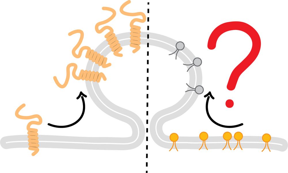
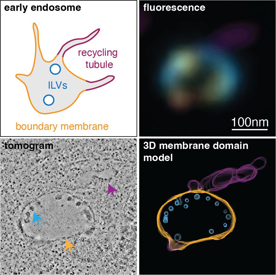
It’s incredibly hard to study lipids in biological membranes on the nanoscale. You need near-perfect information on both membrane ultrastructure and lipid density, which usually isn’t attainable. Lipid-CLEM brought to you by @mathilda95.bsky.social changes that.
www.biorxiv.org/content/10.1...
www.biorxiv.org/content/10.1...
January 31, 2025 at 10:28 AM
It’s incredibly hard to study lipids in biological membranes on the nanoscale. You need near-perfect information on both membrane ultrastructure and lipid density, which usually isn’t attainable. Lipid-CLEM brought to you by @mathilda95.bsky.social changes that.
www.biorxiv.org/content/10.1...
www.biorxiv.org/content/10.1...
Reposted by Milena Schuhmacher
Lipid-CLEM ! Nice, looking forward reading this in depth!

Ooh nice preprint! Studying membrane nanodomains needs nanoscale lipid localization - current methods fall short. 'Lipid-CLEM', correlative light & EM with click chemistry to map lipids. In endosomes, it shows sphingomyelin’s differential partitioning beautifully.
www.biorxiv.org/content/10.1...
www.biorxiv.org/content/10.1...

Visualizing sub-organellar lipid distribution using correlative light and electron microscopy
Lipids and proteins compartmentalize biological membranes into nanoscale domains which are crucial for signaling, intracellular trafficking and many other cellular processes. Studying nanodomain funct...
www.biorxiv.org
January 30, 2025 at 6:22 AM
Lipid-CLEM ! Nice, looking forward reading this in depth!
Reposted by Milena Schuhmacher
Apply now for the ELBE Postdoctoral Fellows Program at the Center for Systems Biology @mpicbg.bsky.social @mpipks.bsky.social. The fully funded fellowships for postdoctoral researchers are designed for 2-3 years. Application deadline is February 14th. Join us! www.csbdresden.de/join-us/jobs...
January 9, 2025 at 1:37 PM
Apply now for the ELBE Postdoctoral Fellows Program at the Center for Systems Biology @mpicbg.bsky.social @mpipks.bsky.social. The fully funded fellowships for postdoctoral researchers are designed for 2-3 years. Application deadline is February 14th. Join us! www.csbdresden.de/join-us/jobs...
Reposted by Milena Schuhmacher
📢📢Official release #PhD position #OrganicChemistry at @Chemie_UMR & @mpi_marburg within Loewe consortium #RobuCop 🤖🦠
Apply online 👇
stellenangebote.uni-marburg.de/jobposting/3...
📆Deadline 22.12 - please RT♥️🙏 #ChemBio
Apply online 👇
stellenangebote.uni-marburg.de/jobposting/3...
📆Deadline 22.12 - please RT♥️🙏 #ChemBio

December 12, 2024 at 7:41 AM
📢📢Official release #PhD position #OrganicChemistry at @Chemie_UMR & @mpi_marburg within Loewe consortium #RobuCop 🤖🦠
Apply online 👇
stellenangebote.uni-marburg.de/jobposting/3...
📆Deadline 22.12 - please RT♥️🙏 #ChemBio
Apply online 👇
stellenangebote.uni-marburg.de/jobposting/3...
📆Deadline 22.12 - please RT♥️🙏 #ChemBio