Alex Ludwig
@alexludwig.bsky.social
1.3K followers
620 following
45 posts
Cell biologist and nature lover interested in epithelial cell junctions, mechanobiology, and cell polarity. Asst Prof at NTU Singapore.
Check out the ALab homepage:
https://blogs.ntu.edu.sg/alabntusg/
Posts
Media
Videos
Starter Packs
Reposted by Alex Ludwig

Hans Clevers
@hansclevers.bsky.social
· Sep 6

Epithelial tension controls intestinal cell extrusion
Cell extrusion is essential for homeostatic self-renewal of the intestinal epithelium. Extrusion is thought to be triggered by crowding-induced compression of cells at the intestinal villus tip. In th...
www.science.org
Reposted by Alex Ludwig

Reposted by Alex Ludwig
Kimberly Kline 🏔
@kimingeneva.bsky.social
· Aug 11

Alex Ludwig
@alexludwig.bsky.social
· Jul 25

Reposted by Alex Ludwig
Felix Campelo
@felixmendu.bsky.social
· Jul 4
Intracellular mechanics and organelle mechanobiology
Mechanobiology is an interdisciplinary field that emerges at the cross-section of biology, physics and engineering. It aims to understand how living cells, tissues and animals sense and respond to me…
meetings.embo.org
Reposted by Alex Ludwig

Alex Ludwig
@alexludwig.bsky.social
· Apr 17
Alex Ludwig
@alexludwig.bsky.social
· Apr 8
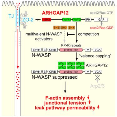
ARHGAP12 suppresses F-actin assembly to control epithelial tight junction mechanics and paracellular leak pathway permeability
Tambrin et al. show that the Cdc42/Rac GAP ARHGAP12 promotes the flux of macromolecules
across tight junctions by suppressing junctional actin assembly and tension via N-WASP
and the Arp2/3 complex. T...
www.cell.com
Alex Ludwig
@alexludwig.bsky.social
· Mar 17
Alex Ludwig
@alexludwig.bsky.social
· Mar 13
Science under siege: protecting scientific progress in turbulent times
As Editors-in-Chief of The Company of Biologists' journals, we have been watching with growing concern the policy changes in the United States of America (USA) and the challenges that these are creati...
journals.biologists.com
Alex Ludwig
@alexludwig.bsky.social
· Mar 7
Alex Ludwig
@alexludwig.bsky.social
· Mar 7

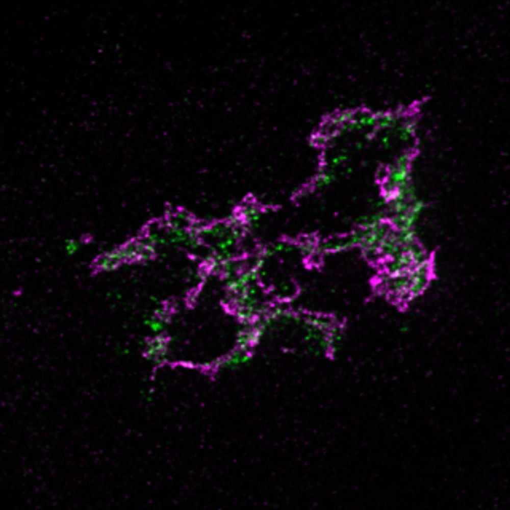
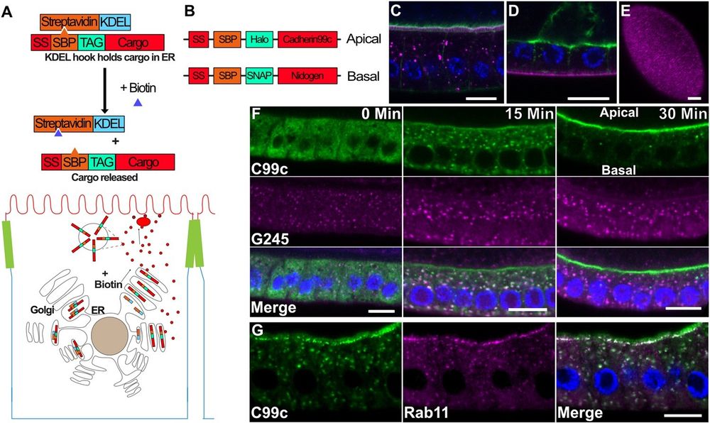
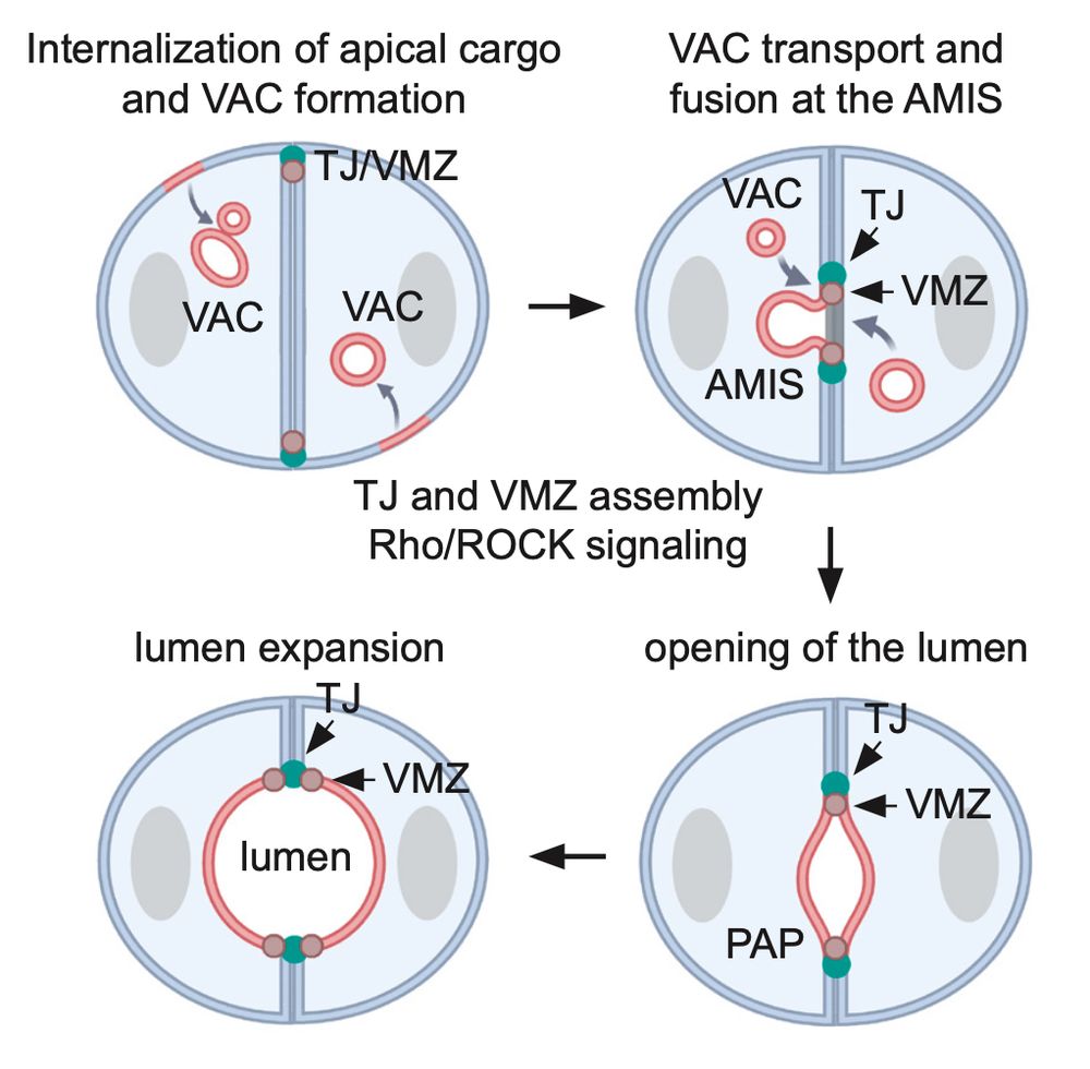
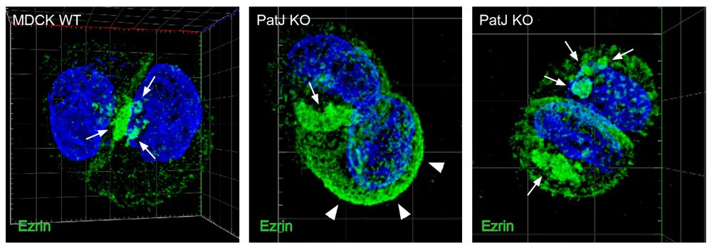
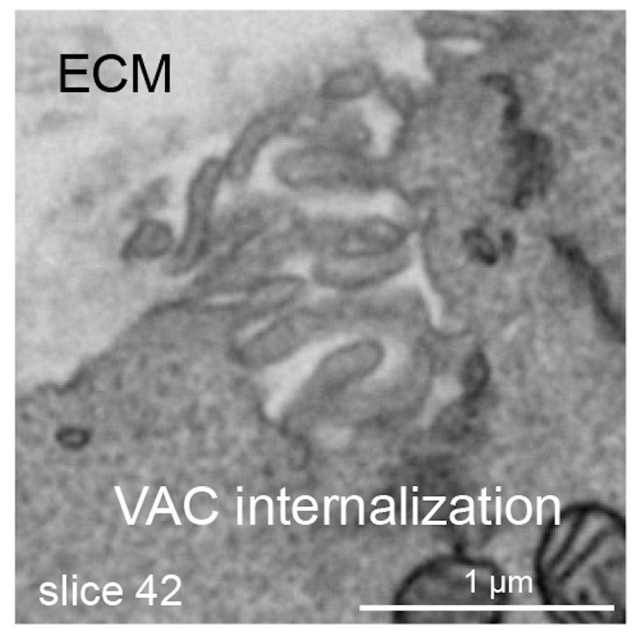
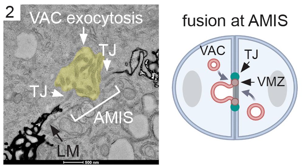
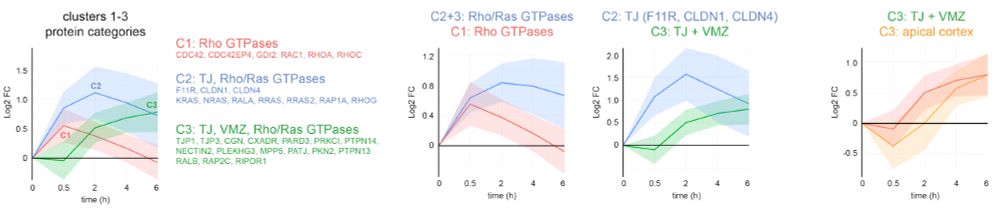
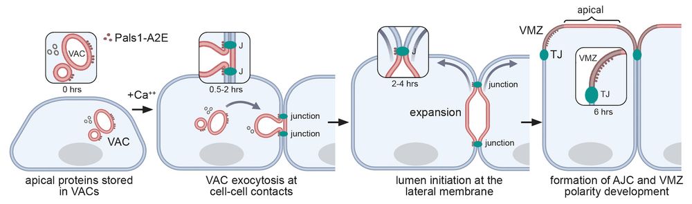
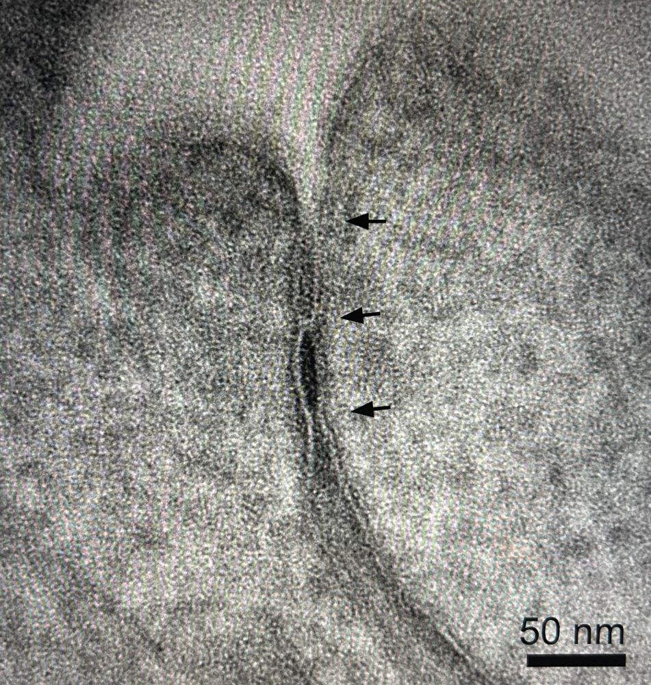
Bulk exocytosis of large intracellular apical precursor organelles establishes apical domain identity during de novo lumen formation
The creation of a microvilli-rich apical luminal domain is a key event in the development of epithelial tissues. De novo lumenogenesis, in which epithelial cells establish apical identity by directing...
www.biorxiv.org
Reposted by Alex Ludwig