
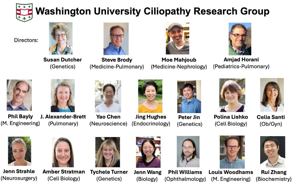
WashU Ciliopathy Research Group
@wu-ciliopathygroup.bsky.social
800 followers
2.7K following
37 posts
A multidisciplinary team of investigators at Washington University in St Louis that perform fundamental, translational, and clinical research on human ciliopathies.
Posts
Media
Videos
Starter Packs
Pinned
Reposted by WashU Ciliopathy Research Group


Reposted by WashU Ciliopathy Research Group

Reposted by WashU Ciliopathy Research Group

Amber Stratman
@astratman.bsky.social
· Jul 15

Reposted by WashU Ciliopathy Research Group
Reposted by WashU Ciliopathy Research Group


Reposted by WashU Ciliopathy Research Group

Reposted by WashU Ciliopathy Research Group


Reposted by WashU Ciliopathy Research Group
I-75 Scientist
@flscitriguy.bsky.social
· Jul 14

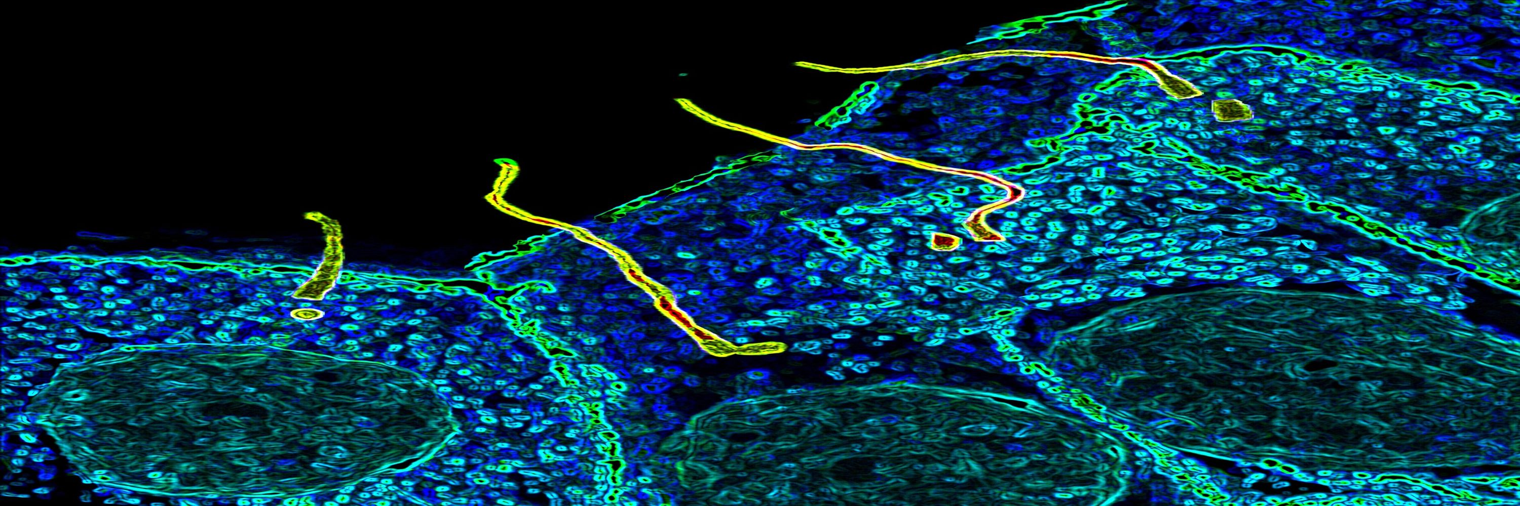
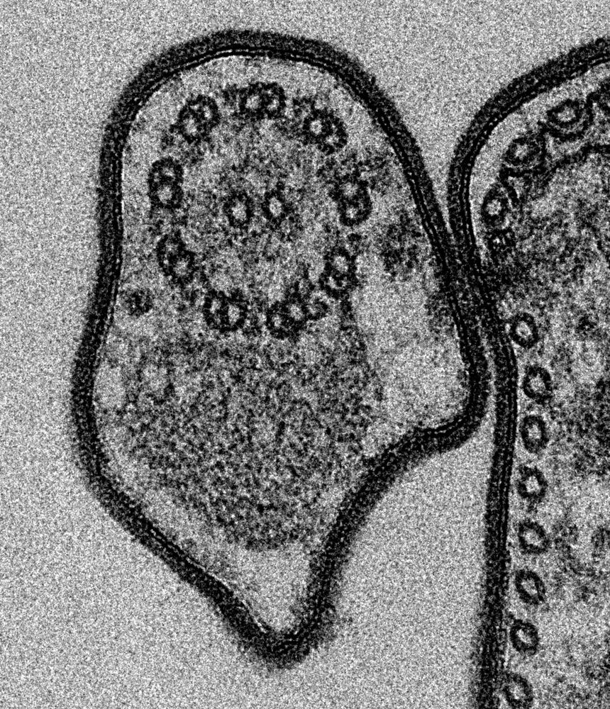
Identification of new ciliary signaling pathways in the brain and insights into neurological disorders
Primary cilia are conserved sensory hubs essential for signaling transduction and embryonic development. Ciliary dysfunction causes a variety of developmental syndromes with neurological features and ...
www.jneurosci.org


Reposted by WashU Ciliopathy Research Group

Reposted by WashU Ciliopathy Research Group


Reposted by WashU Ciliopathy Research Group


Reposted by WashU Ciliopathy Research Group

Reposted by WashU Ciliopathy Research Group
builab.bsky.social
@builab.bsky.social
· Jul 11
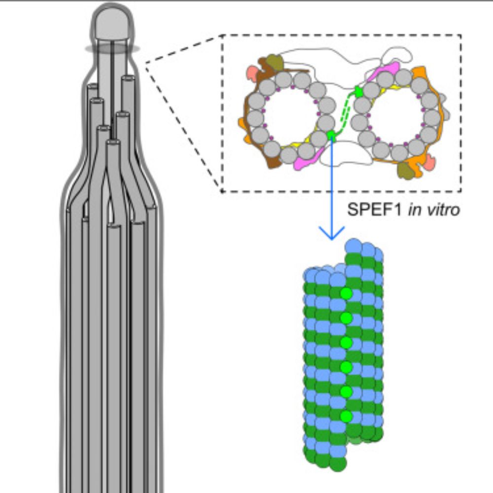
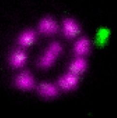
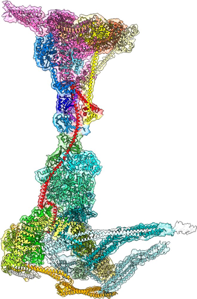
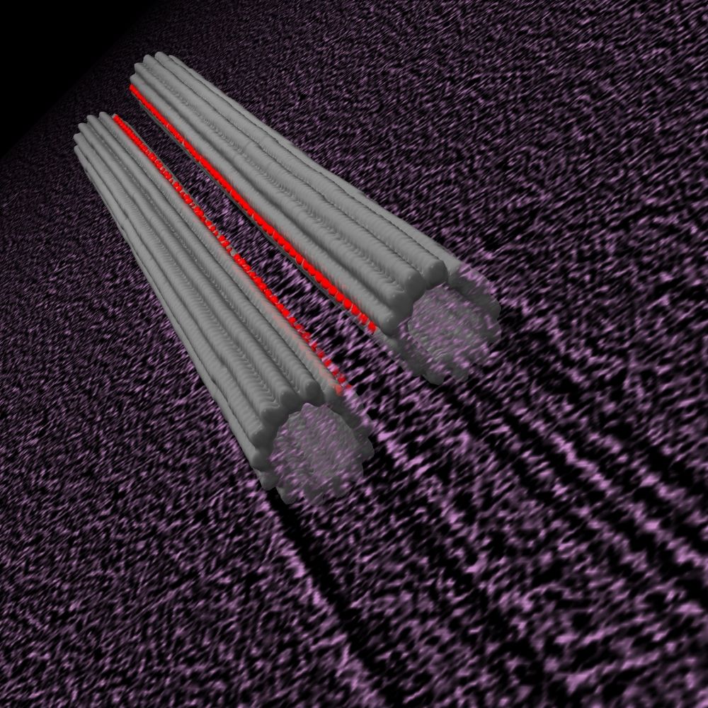
Structure of the ciliary tip central pair reveals the unique role of the microtubule-seam binding protein SPEF1
Motile cilia are unique organelles with the ability to move autonomously. The force generated by beating cilia propels cells and moves fluids. The cil…
www.sciencedirect.com
Reposted by WashU Ciliopathy Research Group

Reposted by WashU Ciliopathy Research Group
Kirsty Wan
@micromotility.bsky.social
· Jul 14
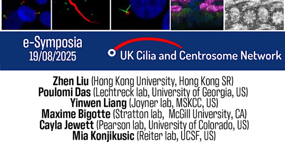
Multi-million project to ‘crack the code’ of cilia – tiny structures with big impact on human health
An international team of researchers, led by the University of Exeter, have been awarded a Wellcome Discovery Award grant of almost £5 million to investigate one of the body’s most fascinating microsc...
news.exeter.ac.uk
Reposted by WashU Ciliopathy Research Group
Reposted by WashU Ciliopathy Research Group


Reposted by WashU Ciliopathy Research Group


Reposted by WashU Ciliopathy Research Group
